1ZZW

Crystal Structure of catalytic domain of Human MAP Kinase Phosphatase 5


Experimental Data Snapshot

  • Method: X-RAY DIFFRACTION
  • Resolution: 1.60 Å
  • R-Value Free: 0.217 
  • R-Value Work: 0.193 
  • R-Value Observed: 0.193 

wwPDB Validation   3D Report Full Report


This is version 1.3 of the entry. See complete history


Literature

Crystal Structure of the Catalytic Domain of Human MAP Kinase Phosphatase 5: Structural Insight into Constitutively Active Phosphatase.

Jeong, D.G.Yoon, T.S.Kim, J.H.Shim, M.Y.Jeong, S.K.Son, J.H.Ryu, S.E.Kim, S.J.

(2006) J Mol Biol 360: 946-955

  • DOI: https://doi.org/10.1016/j.jmb.2006.05.059
  • Primary Citation of Related Structures:  
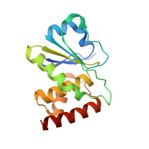
    1ZZW

  • PubMed Abstract: 

    MAP kinase phosphatase 5 (MKP5) is a member of the mitogen-activated protein kinase phosphatase (MKP) family and selectively dephosphorylates JNK and p38. We have determined the crystal structure of the catalytic domain of human MKP5 (MKP5-C) to 1.6 A. In previously reported MKP-C structures, the residues that constitute the active site are seriously deviated from the active conformation of protein tyrosine phosphatases (PTPs), which are accompanied by low catalytic activity. High activities of MKPs are achieved by binding their cognate substrates, representing substrate-induced activation. However, the MKP5-C structure adopts an active conformation of PTP even in the absence of its substrate binding, which is consistent with the previous results that MKP5 solely possesses the intrinsic activity. Further, we identify a sequence motif common to the members of MKPs having low catalytic activity by comparing structures and sequences of other MKPs. Our structural information provides an explanation of constitutive activity of MKP5 as well as the structural insight into substrate-induced activation occurred in other MKPs.


  • Organizational Affiliation

    Systemic Proteomics Research Center, Korea Research Institute of Bioscience and Biotechnology, 52 Eoeun-Dong, Yuseong-Gu, Daejeon, 305-333, Republic of Korea.


Macromolecules
Find similar proteins by:  (by identity cutoff)  |  3D Structure
Entity ID: 1
MoleculeChains Sequence LengthOrganismDetailsImage
Dual specificity protein phosphatase 10
A, B
149Homo sapiensMutation(s): 0 
EC: 3.1.3.48 (PDB Primary Data), 3.1.3.16 (PDB Primary Data)
UniProt & NIH Common Fund Data Resources
Find proteins for Q9Y6W6 (Homo sapiens)
Explore Q9Y6W6 
Go to UniProtKB:  Q9Y6W6
PHAROS:  Q9Y6W6
GTEx:  ENSG00000143507 
Entity Groups  
Sequence Clusters30% Identity50% Identity70% Identity90% Identity95% Identity100% Identity
UniProt GroupQ9Y6W6
Sequence Annotations
Expand
  • Reference Sequence
Experimental Data & Validation

Experimental Data

  • Method: X-RAY DIFFRACTION
  • Resolution: 1.60 Å
  • R-Value Free: 0.217 
  • R-Value Work: 0.193 
  • R-Value Observed: 0.193 
  • Space Group: P 1
Unit Cell:
Length ( Å )Angle ( ˚ )
a = 39.533α = 95.35
b = 41.073β = 99.81
c = 55.569γ = 117.35
Software Package:
Software NamePurpose
MOSFLMdata reduction
SCALAdata scaling
EPMRphasing
CNSrefinement
CCP4data scaling

Structure Validation

View Full Validation Report


Entry History 

Deposition Data

Revision History  (Full details and data files)

  • Version 1.0: 2006-07-04
    Type: Initial release
  • Version 1.1: 2008-04-30
    Changes: Version format compliance
  • Version 1.2: 2011-07-13
    Changes: Version format compliance
  • Version 1.3: 2024-03-13
    Changes: Data collection, Database references, Derived calculations