1Z9S

Crystal Structure of the native chaperone:subunit:subunit Caf1M:Caf1:Caf1 complex


Experimental Data Snapshot

  • Method: X-RAY DIFFRACTION
  • Resolution: 2.20 Å
  • R-Value Free: 0.243 
  • R-Value Work: 0.223 
  • R-Value Observed: 0.224 

wwPDB Validation   3D Report Full Report


This is version 1.2 of the entry. See complete history


Literature

Resolving the energy paradox of chaperone/usher-mediated fibre assembly

Zavialov, A.V.Tischenko, V.M.Fooks, L.J.Brandsdal, B.O.Aqvist, J.Zav'yalov, V.P.Macintyre, S.Knight, S.D.

(2005) Biochem J 389: 685-694

  • DOI: https://doi.org/10.1042/BJ20050426
  • Primary Citation of Related Structures:  
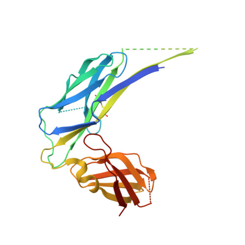
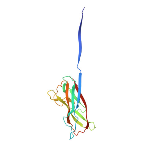
    1Z9S

  • PubMed Abstract: 

    Periplasmic chaperone/usher machineries are used for assembly of filamentous adhesion organelles of Gram-negative pathogens in a process that has been suggested to be driven by folding energy. Structures of mutant chaperone-subunit complexes revealed a final folding transition (condensation of the subunit hydrophobic core) on the release of organelle subunit from the chaperone-subunit pre-assembly complex and incorporation into the final fibre structure. However, in view of the large interface between chaperone and subunit in the pre-assembly complex and the reported stability of this complex, it is difficult to understand how final folding could release sufficient energy to drive assembly. In the present paper, we show the X-ray structure for a native chaperone-fibre complex that, together with thermodynamic data, shows that the final folding step is indeed an essential component of the assembly process. We show that completion of the hydrophobic core and incorporation into the fibre results in an exceptionally stable module, whereas the chaperone-subunit pre-assembly complex is greatly destabilized by the high-energy conformation of the bound subunit. This difference in stabilities creates a free energy potential that drives fibre formation.


  • Organizational Affiliation

    Department of Molecular Biology, Uppsala Biomedical Center, Swedish University of Agricultural Sciences, Box 590, SE-753 24 Uppsala, Sweden. anton.zavialov@molbio.slu.se


Macromolecules
Find similar proteins by:  (by identity cutoff)  |  3D Structure
Entity ID: 1
MoleculeChains Sequence LengthOrganismDetailsImage
Chaperone protein Caf1M235Yersinia pestisMutation(s): 0 
Gene Names: caf1M
UniProt
Find proteins for P26926 (Yersinia pestis)
Explore P26926 
Go to UniProtKB:  P26926
Entity Groups  
Sequence Clusters30% Identity50% Identity70% Identity90% Identity95% Identity100% Identity
UniProt GroupP26926
Sequence Annotations
Expand
  • Reference Sequence
Find similar proteins by:  (by identity cutoff)  |  3D Structure
Entity ID: 2
MoleculeChains Sequence LengthOrganismDetailsImage
F1 capsule antigen
B, C
149Yersinia pestisMutation(s): 0 
Gene Names: caf1
UniProt
Find proteins for P26948 (Yersinia pestis)
Explore P26948 
Go to UniProtKB:  P26948
Entity Groups  
Sequence Clusters30% Identity50% Identity70% Identity90% Identity95% Identity100% Identity
UniProt GroupP26948
Sequence Annotations
Expand
  • Reference Sequence
Experimental Data & Validation

Experimental Data

  • Method: X-RAY DIFFRACTION
  • Resolution: 2.20 Å
  • R-Value Free: 0.243 
  • R-Value Work: 0.223 
  • R-Value Observed: 0.224 
  • Space Group: P 21 21 2
Unit Cell:
Length ( Å )Angle ( ˚ )
a = 69.8α = 90
b = 180.14β = 90
c = 45.73γ = 90
Software Package:
Software NamePurpose
MOSFLMdata reduction
SCALAdata scaling
MOLREPphasing
CNSrefinement
CCP4data scaling

Structure Validation

View Full Validation Report


Entry History 

Deposition Data

Revision History  (Full details and data files)

  • Version 1.0: 2005-06-21
    Type: Initial release
  • Version 1.1: 2008-04-30
    Changes: Version format compliance
  • Version 1.2: 2011-07-13
    Changes: Version format compliance