A Structural Model for the Membrane-bound Form of the Juxtamembrane Domain of the Epidermal Growth Factor Receptor
Choowongkomon, K., Carlin, C.R.(2005) J Biol Chem 280: 24043-24052
- PubMed: 15840573
- DOI: https://doi.org/10.1074/jbc.M502698200
- Primary Citation of Related Structures:
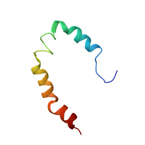
1Z9I - PubMed Abstract:
The epidermal growth factor receptor (EGFR) is a member of the receptor tyrosine kinase family involved in the regulation of cellular proliferation and differentiation. Its juxtamembrane domain (JX), the region located between the transmembrane and kinase domains, plays important roles in receptor trafficking. Two sorting signals, a PXXP motif and a 658LL659 motif, are responsible for basolateral sorting in polarized epithelial cells, and a 679LL680 motif targets the ligand-activated receptor for lysosomal degradation. To understand the regulation of these signals, we characterized the structural properties of recombinant JX domain in aqueous solution and in dodecylphosphocholine (DPC) detergent. JX is inherently unstructured in aqueous solution, albeit a nascent helix encompasses the lysosomal sorting signal. In DPC micelles, structures derived from NMR data showed three amphipathic, helical segments. A large, internally inconsistent group of long range nuclear Overhauser effects suggest a close proximity of the helices, and the presence of significant conformational averaging. Models were determined for the average JX conformation using restraints representing the translational restriction due to micelle-surface adsorption, and the helix orientations were determined from residual dipolar couplings. Two equivalent average structural models were obtained that differ only in the relative orientation between first and second helices. In these models, the 658LL659 and 679LL680 motifs are located in the first and second helices and face the micelle surface, whereas the PXXP motif is located in a flexible helix-connecting region. The data suggest that the activity of these signals may be regulated by their membrane association and restricted accessibility in the intact receptor.
Organizational Affiliation:
Department of Physiology and Biophysics, School of Medicine, Case Western Reserve University, Cleveland, OH 44106-4970, USA.