1Z6F

Crystal structure of penicillin-binding protein 5 from E. coli in complex with a boronic acid inhibitor


Experimental Data Snapshot

  • Method: X-RAY DIFFRACTION
  • Resolution: 1.60 Å
  • R-Value Free: 0.240 
  • R-Value Work: 0.213 
  • R-Value Observed: 0.215 

wwPDB Validation   3D Report Full Report


Ligand Structure Quality Assessment 


This is version 1.4 of the entry. See complete history


Literature

Crystal Structure of Escherichia coli Penicillin-Binding Protein 5 Bound to a Tripeptide Boronic Acid Inhibitor: A Role for Ser-110 in Deacylation.

Nicola, G.Peddi, S.Stefanova, M.Nicholas, R.A.Gutheil, W.G.Davies, C.

(2005) Biochemistry 44: 8207-8217

  • DOI: https://doi.org/10.1021/bi0473004
  • Primary Citation of Related Structures:  
    1Z6F

  • PubMed Abstract: 

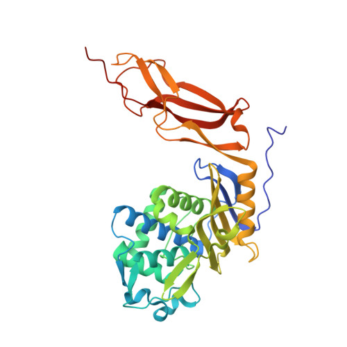
    Penicillin-binding protein 5 (PBP 5) from Escherichia coli is a well-characterized d-alanine carboxypeptidase that serves as a prototypical enzyme to elucidate the structure, function, and catalytic mechanism of PBPs. A comprehensive understanding of the catalytic mechanism underlying d-alanine carboxypeptidation and antibiotic binding has proven elusive. In this study, we report the crystal structure at 1.6 A resolution of PBP 5 in complex with a substrate-like peptide boronic acid, which was designed to resemble the transition-state intermediate during the deacylation step of the enzyme-catalyzed reaction with peptide substrates. In the structure of the complex, the boron atom is covalently attached to Ser-44, which in turn is within hydrogen-bonding distance to Lys-47. This arrangement further supports the assignment of Lys-47 as the general base that activates Ser-44 during acylation. One of the two hydroxyls in the boronyl center (O2) is held by the oxyanion hole comprising the amides of Ser-44 and His-216, while the other hydroxyl (O3), which is analogous to the nucleophilic water for hydrolysis of the acyl-enzyme intermediate, is solvated by a water molecule that bridges to Ser-110. Lys-47 is not well-positioned to act as the catalytic base in the deacylation reaction. Instead, these data suggest a mechanism of catalysis for deacylation that uses a hydrogen-bonding network, involving Lys-213, Ser-110, and a bridging water molecule, to polarize the hydrolytic water molecule.


  • Organizational Affiliation

    Department of Biochemistry, Medical University of South Carolina, Charleston, South Carolina 29425, USA.


Macromolecules
Find similar proteins by:  (by identity cutoff)  |  3D Structure
Entity ID: 1
MoleculeChains Sequence LengthOrganismDetailsImage
Penicillin-binding protein 5363Escherichia coliMutation(s): 0 
Gene Names: dacApfv
EC: 3.4.16.4
UniProt
Find proteins for P0AEB2 (Escherichia coli (strain K12))
Explore P0AEB2 
Go to UniProtKB:  P0AEB2
Entity Groups  
Sequence Clusters30% Identity50% Identity70% Identity90% Identity95% Identity100% Identity
UniProt GroupP0AEB2
Sequence Annotations
Expand
  • Reference Sequence
Small Molecules
Ligands 2 Unique
IDChains Name / Formula / InChI Key2D Diagram3D Interactions
BO9
Query on BO9

Download Ideal Coordinates CCD File 
B [auth A]N1-[(1R)-1-(DIHYDROXYBORYL)ETHYL]-N2-[(TERT-BUTOXYCARBONYL)-D-GAMMA-GLUTAMYL]-N6-[(BENZYLOXY)CARBONYL-L-LYSINAMIDE
C26 H41 B N4 O10
OTTYWVUOPWENAJ-HOJAQTOUSA-N
GOL
Query on GOL

Download Ideal Coordinates CCD File 
C [auth A]GLYCEROL
C3 H8 O3
PEDCQBHIVMGVHV-UHFFFAOYSA-N
Binding Affinity Annotations 
IDSourceBinding Affinity
BO9 PDBBind:  1Z6F Ki: 1.30e+4 (nM) from 1 assay(s)
Experimental Data & Validation

Experimental Data

  • Method: X-RAY DIFFRACTION
  • Resolution: 1.60 Å
  • R-Value Free: 0.240 
  • R-Value Work: 0.213 
  • R-Value Observed: 0.215 
  • Space Group: C 1 2 1
Unit Cell:
Length ( Å )Angle ( ˚ )
a = 109.76α = 90
b = 50.25β = 120.2
c = 84.31γ = 90
Software Package:
Software NamePurpose
REFMACrefinement
MAR345data collection
SCALEPACKdata scaling

Structure Validation

View Full Validation Report


Ligand Structure Quality Assessment 


Entry History 

Deposition Data

Revision History  (Full details and data files)

  • Version 1.0: 2005-06-21
    Type: Initial release
  • Version 1.1: 2008-04-30
    Changes: Version format compliance
  • Version 1.2: 2011-07-13
    Changes: Non-polymer description, Version format compliance
  • Version 1.3: 2017-10-11
    Changes: Advisory, Refinement description
  • Version 1.4: 2023-08-23
    Changes: Advisory, Data collection, Database references, Derived calculations, Refinement description