1Z5T

Crystal Structure of [d(CGCGAA(Z3dU)(Z3dU)CGCG)]2, Z3dU:5-(3-aminopropyl)-2'-deoxyuridine, in presence of thallium I.


Experimental Data Snapshot

  • Method: X-RAY DIFFRACTION
  • Resolution: 1.60 Å
  • R-Value Free: 0.244 
  • R-Value Work: 0.198 
  • R-Value Observed: 0.233 

wwPDB Validation   3D Report Full Report


Ligand Structure Quality Assessment 


This is version 1.5 of the entry. See complete history


Literature

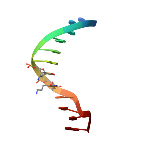
Structure of B-DNA with cations tethered in the major groove.

Moulaei, T.Maehigashi, T.Lountos, G.T.Komeda, S.Watkins, D.Stone, M.P.Marky, L.A.Li, J.S.Gold, B.Williams, L.D.

(2005) Biochemistry 44: 7458-7468

  • DOI: https://doi.org/10.1021/bi050128z
  • Primary Citation of Related Structures:  
    1Z5T

  • PubMed Abstract: 

    Here, we describe the 1.6-A X-ray structure of the DDD (Dickerson-Drew dodecamer), which has been covalently modified by the tethering of four cationic charges. This modified version of the DDD, called here the DDD(4+), is composed of [d(CGCGAAXXCGCG)](2), where X is effectively a thymine residue linked at the 5 position to an n-propyl-amine. The structure was determined from crystals soaked with thallium(I), which has been broadly used as a mimic of K(+) in X-ray diffraction experiments aimed at determining positions of cations adjacent to nucleic acids. Three of the tethered cations are directed radially out from the DNA. The radially directed tethered cations do not appear to induce structural changes or to displace counterions. One of the tethered cations is directed in the 3' direction, toward a phosphate group near one end of the duplex. This tethered cation appears to interact electrostatically with the DNA. This interaction is accompanied by changes in helical parameters rise, roll, and twist and by a displacement of the backbone relative to a control oligonucleotide. In addition, these interactions appear to be associated with displacement of counterions from the major groove of the DNA.


  • Organizational Affiliation

    Department of Chemistry and Biochemistry, Georgia Institute of Technology, Atlanta, Georgia 30332-0400, USA.


Macromolecules

Find similar nucleic acids by:  Sequence   |   3D Structure  

Entity ID: 1
MoleculeChains LengthOrganismImage
5'-D(*CP*GP*CP*GP*AP*AP*(ZDU)P*(ZDU)P*CP*GP*CP*G)-3'
A, B
12N/A
Sequence Annotations
Expand
  • Reference Sequence
Small Molecules
Ligands 2 Unique
IDChains Name / Formula / InChI Key2D Diagram3D Interactions
TL
Query on TL

Download Ideal Coordinates CCD File 
D [auth A]
E [auth A]
F [auth A]
G [auth A]
H [auth A]
D [auth A],
E [auth A],
F [auth A],
G [auth A],
H [auth A],
J [auth B],
K [auth B],
L [auth B]
THALLIUM (I) ION
Tl
ZLUSCZLCHQSJRU-UHFFFAOYSA-N
SPM
Query on SPM

Download Ideal Coordinates CCD File 
C [auth A],
I [auth B]
SPERMINE
C10 H26 N4
PFNFFQXMRSDOHW-UHFFFAOYSA-N
Experimental Data & Validation

Experimental Data

  • Method: X-RAY DIFFRACTION
  • Resolution: 1.60 Å
  • R-Value Free: 0.244 
  • R-Value Work: 0.198 
  • R-Value Observed: 0.233 
  • Space Group: P 21 21 21
Unit Cell:
Length ( Å )Angle ( ˚ )
a = 24.994α = 90
b = 41.057β = 90
c = 66.339γ = 90
Software Package:
Software NamePurpose
DENZOdata reduction
SCALEPACKdata scaling
CNSrefinement
HKL-2000data reduction
CNSphasing

Structure Validation

View Full Validation Report


Ligand Structure Quality Assessment 


Entry History 

Deposition Data

Revision History  (Full details and data files)

  • Version 1.0: 2005-07-26
    Type: Initial release
  • Version 1.1: 2008-04-30
    Changes: Version format compliance
  • Version 1.2: 2011-07-13
    Changes: Version format compliance
  • Version 1.3: 2017-10-11
    Changes: Advisory, Refinement description
  • Version 1.4: 2024-02-14
    Changes: Advisory, Data collection, Database references, Derived calculations
  • Version 1.5: 2024-04-03
    Changes: Refinement description