The Modular Structure of SIP Facilitates Its Role in Stabilizing Multiprotein Assemblies.
Bhattacharya, S., Lee, Y.T., Michowski, W., Jastrzebska, B., Filipek, A., Kuznicki, J., Chazin, W.J.(2005) Biochemistry 44: 9462-9471
- PubMed: 15996101
- DOI: https://doi.org/10.1021/bi0502689
- Primary Citation of Related Structures:
1YSM - PubMed Abstract:
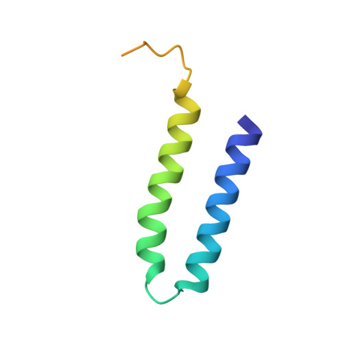
Siah-interacting protein (SIP) was identified as a novel adaptor that physically links the E3 ubiquitin ligase activity of Siah-1 with Skp1 and Ebi F-Box protein in the degradation of beta-catenin, a transcriptional activator of TCF/LEF genes. In this study, we have used solution NMR spectroscopy to characterize the domain structure of SIP, which includes a novel helical hairpin domain at the N-terminus flexibly linked to a CS domain and an unstructured carboxy terminal SGS domain. These studies have been complemented by mapping the sites of functionally important protein-protein interactions involving Siah-1 and Skp1 to individual domains of SIP. NMR-based chemical shift perturbation assays show that Siah-1 interacts with the flexible linker between SIP N and CS domains. This site for interaction in the linker does not perturb residues in the structured region at the N-terminus but does appear to restrict the rotational freedom of the SIP CS domain in the context of the full-length protein. In contrast, Skp1 engages the SIP CS domain exclusively through weak interactions that are not coupled to the other domains. The principal role of the modular structure of SIP appears to be in bringing these two proteins into physical proximity and orchestrating the orientation required for polyubiquitination of beta-catenin in the intact SCF-type complex.
Organizational Affiliation:
Department of Biochemistry, Center for Structural Biology, 5140 BIOSCI/MRBIII, Vanderbilt University, Nashville, Tennessee 37232-8725, USA.