The three-dimensional structure of two mutants of the signal transduction protein CheY suggest its molecular activation mechanism.
Bellsolell, L., Cronet, P., Majolero, M., Serrano, L., Coll, M.(1996) J Mol Biol 257: 116-128
- PubMed: 8632450
- DOI: https://doi.org/10.1006/jmbi.1996.0151
- Primary Citation of Related Structures:
1YMU, 1YMV - PubMed Abstract:
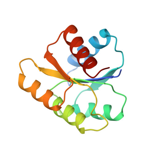
The three-dimensional crystal structures of the single mutant M17G and the triple mutant F14G-S15G-M17G of the response regulator protein CheY have been determined to 2.3 and 1.9 angstrom, respectively. Both mutants bind the essential Mg2+ cation as determined by the changes in stability, but binding does not cause the intrinsic fluorescence quenching of W58 observed in the wild-type protein. The loop beta4-alpha4 appears to be very flexible in both mutants and helix alpha4, which starts at N94 in the native Mg2+-CheY and at K91 in the native apo-CheY, starts in both mutants at residue K92. The side-chain of K109 appears to be more mobile because of the space freed by the M17G mutation. In the triple mutant the main chain of K109 and adjacent residues (loop beta5-alpha5) is displaced almost by 2 angstrom affecting the main chain at residues T87 to E89 (C terminus of beta4). The triple mutant structure has a Mg2+ bound at the active site, but although the Mg2+ coordination is similar to that of the native Mg2+-CheY, the structural consequences of the metal binding are quite different. It seems that the mutations have disrupted the mechanism of movement transmission observed in the native protein. We suggest that the side-chain of K109, packed between V86, A88 and M17 in the native protein, slides forwards and backwards upon activation and deactivation dragging the main chain at the loop beta5-alpha5 and triggering larger movements at the functional surface of the protein.
Organizational Affiliation:
Departament de Biologia Molecular i Cel-lular Centre d'Investigacio i Desenvolupament-CSIC, Barcelona, Spain.