1Y6O

Crystal structure of disulfide engineered porcine pancreatic phospholipase A2 to group-X isozyme in complex with inhibitor MJ33 and phosphate ions


Experimental Data Snapshot

  • Method: X-RAY DIFFRACTION
  • Resolution: 2.00 Å
  • R-Value Free: 0.245 
  • R-Value Work: 0.196 
  • R-Value Observed: 0.196 

wwPDB Validation   3D Report Full Report


Ligand Structure Quality Assessment 


This is version 1.4 of the entry. See complete history


Literature

Kinetic and structural properties of disulfide engineered phospholipase a(2): insight into the role of disulfide bonding patterns.

Yu, B.Z.Pan, Y.H.Janssen, M.J.W.Bahnson, B.J.Jain, M.K.

(2005) Biochemistry 44: 3369-3379

  • DOI: https://doi.org/10.1021/bi0482147
  • Primary Citation of Related Structures:  
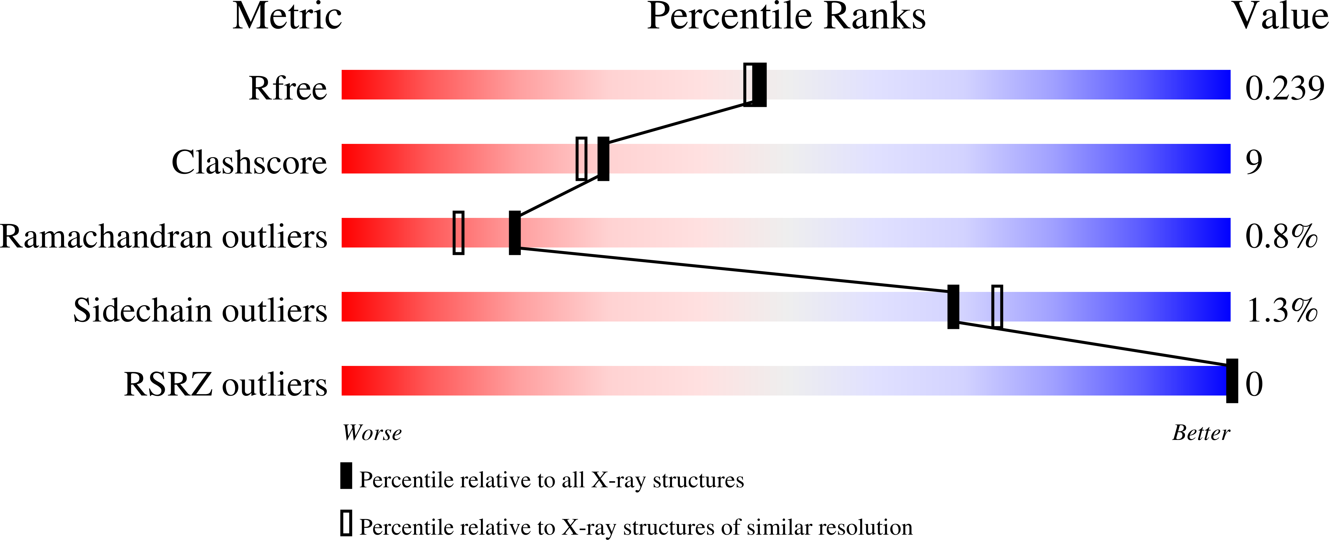
    1Y6O, 1Y6P

  • PubMed Abstract: 

    The family of secreted 14 kDa phospholipase A(2) (PLA2) enzymes have a common motif for the catalytic site but differ in their disulfide architecture. The functional significance of such structural changes has been analyzed by comparing the kinetic and spectroscopic properties of a series of disulfide mutants engineered into the sequence of pig pancreatic IB PLA2 to resemble the mammalian paralogues of the PLA2 family [Janssen et al. (1999) Eur. J. Biochem. 261, 197-207, 1999]. We report a detailed comparison of the functional parameters of pig iso-PLA2, as well as several of the human homologues, with these disulfide engineered mutants of pig IB PLA2. The crystal structure of the ligand free and the active site inhibitor-MJ33 bound forms of PLA2 engineered to have the disulfide bonding pattern of group-X (eng-X) are also reported and compared with the structure of group-IB and human group-X PLA2. The engineered mutants show noticeable functional differences that are rationalized in terms of spectroscopic properties and the differences detected in the crystal structure of eng-X. A major difference between the eng-mutants is in the calcium binding to the enzyme in the aqueous phase, which also influences the binding of the active site directed ligands. We suggest that the disulfide architecture of the PLA2 paralogues has a marginal influence on interface binding. In this comparison, the modest differences observed in the interfacial kinetics are attributed to the changes in the side chain residues. This in turn influences the coupling of the catalytic cycle to the calcium binding and the interfacial binding event.


  • Organizational Affiliation

    Department of Chemistry & Biochemistry, University of Delaware, Newark, Delaware 19716, USA.


Macromolecules
Find similar proteins by:  (by identity cutoff)  |  3D Structure
Entity ID: 1
MoleculeChains Sequence LengthOrganismDetailsImage
Phospholipase A2, major isoenzyme
A, B
131Sus scrofaMutation(s): 3 
Gene Names: PLA2G1B
EC: 3.1.1.4
UniProt
Find proteins for P00592 (Sus scrofa)
Explore P00592 
Go to UniProtKB:  P00592
Entity Groups  
Sequence Clusters30% Identity50% Identity70% Identity90% Identity95% Identity100% Identity
UniProt GroupP00592
Sequence Annotations
Expand
  • Reference Sequence
Small Molecules
Ligands 3 Unique
IDChains Name / Formula / InChI Key2D Diagram3D Interactions
MJI
Query on MJI

Download Ideal Coordinates CCD File 
I [auth A],
N [auth B]
1-HEXADECYL-3-TRIFLUOROETHYL-SN-GLYCERO-2-PHOSPHATE METHANE
C22 H44 F3 O6 P
XPTFBVFCGHXMRK-OAQYLSRUSA-N
PO4
Query on PO4

Download Ideal Coordinates CCD File 
D [auth A]
E [auth A]
F [auth A]
G [auth A]
H [auth A]
D [auth A],
E [auth A],
F [auth A],
G [auth A],
H [auth A],
K [auth B],
L [auth B],
M [auth B]
PHOSPHATE ION
O4 P
NBIIXXVUZAFLBC-UHFFFAOYSA-K
CA
Query on CA

Download Ideal Coordinates CCD File 
C [auth A],
J [auth B]
CALCIUM ION
Ca
BHPQYMZQTOCNFJ-UHFFFAOYSA-N
Experimental Data & Validation

Experimental Data

  • Method: X-RAY DIFFRACTION
  • Resolution: 2.00 Å
  • R-Value Free: 0.245 
  • R-Value Work: 0.196 
  • R-Value Observed: 0.196 
  • Space Group: P 1 21 1
Unit Cell:
Length ( Å )Angle ( ˚ )
a = 38.534α = 90
b = 85.921β = 94.76
c = 38.554γ = 90
Software Package:
Software NamePurpose
CNSrefinement
DENZOdata reduction
SCALEPACKdata scaling
AMoREphasing

Structure Validation

View Full Validation Report


Ligand Structure Quality Assessment 


Entry History 

Deposition Data

Revision History  (Full details and data files)

  • Version 1.0: 2005-03-22
    Type: Initial release
  • Version 1.1: 2008-04-30
    Changes: Version format compliance
  • Version 1.2: 2011-07-13
    Changes: Derived calculations, Version format compliance
  • Version 1.3: 2021-10-20
    Changes: Database references, Derived calculations
  • Version 1.4: 2023-08-23
    Changes: Data collection, Refinement description