1WUY

Crystallographic studies on acyl ureas, a new class of inhibitors of glycogen phosphorylase. Broad specificity of the allosteric site


Experimental Data Snapshot

  • Method: X-RAY DIFFRACTION
  • Resolution: 2.26 Å
  • R-Value Free: 0.232 
  • R-Value Work: 0.191 
  • R-Value Observed: 0.191 

wwPDB Validation   3D Report Full Report


This is version 1.2 of the entry. See complete history


Literature

Crystallographic studies on acyl ureas, a new class of glycogen phosphorylase inhibitors, as potential antidiabetic drugs

Oikonomakos, N.G.Kosmopoulou, M.N.Chrysina, E.D.Leonidas, D.D.Kostas, I.D.Wendt, K.U.Klabunde, T.Defossa, E.

(2005) Protein Sci 14: 1760-1771

  • DOI: https://doi.org/10.1110/ps.051432405
  • Primary Citation of Related Structures:  
    1WUT, 1WUY, 1WV0, 1WV1

  • PubMed Abstract: 

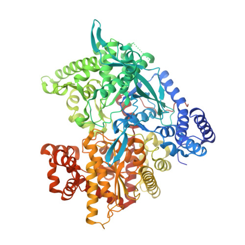
    Acyl ureas were discovered as a novel class of inhibitors for glycogen phosphorylase, a molecular target to control hyperglycemia in type 2 diabetics. This series is exemplified by 6-{2,6-Dichloro- 4-[3-(2-chloro-benzoyl)-ureido]-phenoxy}-hexanoic acid, which inhibits human liver glycogen phosphorylase a with an IC(50) of 2.0 microM. Here we analyze four crystal structures of acyl urea derivatives in complex with rabbit muscle glycogen phosphorylase b to elucidate the mechanism of inhibition of these inhibitors. The structures were determined and refined to 2.26 Angstroms resolution and demonstrate that the inhibitors bind at the allosteric activator site, where the physiological activator AMP binds. Acyl ureas induce conformational changes in the vicinity of the allosteric site. Our findings suggest that acyl ureas inhibit glycogen phosphorylase by direct inhibition of AMP binding and by indirect inhibition of substrate binding through stabilization of the T' state.


  • Organizational Affiliation

    Institute of Organic and Pharmaceutical Chemistry, The National Hellenic Research Foundation, Athens, Greece. ngo@eie.gr


Macromolecules
Find similar proteins by:  (by identity cutoff)  |  3D Structure
Entity ID: 1
MoleculeChains Sequence LengthOrganismDetailsImage
Glycogen phosphorylase, muscle form842Oryctolagus cuniculusMutation(s): 0 
EC: 2.4.1.1
UniProt
Find proteins for P00489 (Oryctolagus cuniculus)
Explore P00489 
Go to UniProtKB:  P00489
Entity Groups  
Sequence Clusters30% Identity50% Identity70% Identity90% Identity95% Identity100% Identity
UniProt GroupP00489
Sequence Annotations
Expand
  • Reference Sequence
Small Molecules
Ligands 2 Unique
IDChains Name / Formula / InChI Key2D Diagram3D Interactions
BN3
Query on BN3

Download Ideal Coordinates CCD File 
C [auth A]4-[3-CHLORO-4-({[(2,4-DICHLOROBENZOYL)AMINO]CARBONYL}AMINO)PHENOXY]BUTANOIC ACID
C18 H15 Cl3 N2 O5
FYQVFMLCZJZZEM-UHFFFAOYSA-N
PLP
Query on PLP

Download Ideal Coordinates CCD File 
B [auth A]PYRIDOXAL-5'-PHOSPHATE
C8 H10 N O6 P
NGVDGCNFYWLIFO-UHFFFAOYSA-N
Binding Affinity Annotations 
IDSourceBinding Affinity
BN3 Binding MOAD:  1WUY IC50: 1600 (nM) from 1 assay(s)
Experimental Data & Validation

Experimental Data

  • Method: X-RAY DIFFRACTION
  • Resolution: 2.26 Å
  • R-Value Free: 0.232 
  • R-Value Work: 0.191 
  • R-Value Observed: 0.191 
  • Space Group: P 43 21 2
Unit Cell:
Length ( Å )Angle ( ˚ )
a = 128.578α = 90
b = 128.578β = 90
c = 116.043γ = 90
Software Package:
Software NamePurpose
X-PLORrefinement
HKL-2000data reduction
SCALEPACKdata scaling
X-PLORphasing

Structure Validation

View Full Validation Report


Entry History 

Deposition Data

Revision History  (Full details and data files)

  • Version 1.0: 2005-12-09
    Type: Initial release
  • Version 1.1: 2008-04-30
    Changes: Version format compliance
  • Version 1.2: 2011-07-13
    Changes: Derived calculations, Version format compliance