1WTD

Crystal structure of type II restrcition endonuclease, EcoO109I DNA-free form


Experimental Data Snapshot

  • Method: X-RAY DIFFRACTION
  • Resolution: 2.40 Å
  • R-Value Free: 0.297 
  • R-Value Work: 0.255 
  • R-Value Observed: 0.257 

wwPDB Validation   3D Report Full Report


This is version 1.3 of the entry. See complete history


Literature

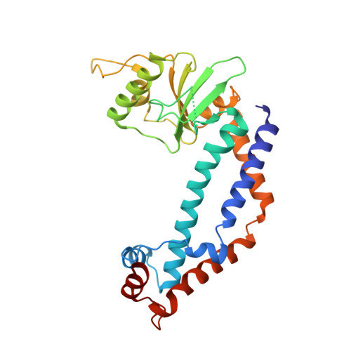
Crystal structures of type II restriction endonuclease EcoO109I and its complex with cognate DNA

Hashimoto, H.Shimizu, T.Imasaki, T.Kato, M.Shichijo, N.Kita, K.Sato, M.

(2005) J Biol Chem 280: 5605-5610

  • DOI: https://doi.org/10.1074/jbc.M411684200
  • Primary Citation of Related Structures:  
    1WTD, 1WTE

  • PubMed Abstract: 

    EcoO109I is a type II restriction endonuclease that recognizes the DNA sequence of RGGNCCY. Here we describe the crystal structures of EcoO109I and its complex with DNA. A comparison of the two structures shows that the catalytic domain moves drastically to capture the DNA. One metal ion and two water molecules are observed near the active site of the DNA complex. The metal ion is a Lewis acid that stabilizes the pentavalent phosphorus atom in the transition state. One water molecule, activated by Lys-126, attacks the phosphorus atom in an S(N)2 mechanism, whereas the other water interacts with the 3'-leaving oxygen to donate a proton to the oxygen. EcoO109I is similar to EcoRI family enzymes in terms of its DNA cleavage pattern and folding topology of the common motif in the catalytic domain, but it differs in the manner of DNA recognition. Our findings propose a novel classification of the type II restriction endonucleases and lead to the suggestion that EcoO109I represents a new subclass of the EcoRI family.


  • Organizational Affiliation

    Graduate School of Integrated Science, Yokohama City University, 1-7-29 Suehiro-cho, Tsurumi-ku, Yokohama 230-0045, Japan.


Macromolecules
Find similar proteins by:  (by identity cutoff)  |  3D Structure
Entity ID: 1
MoleculeChains Sequence LengthOrganismDetailsImage
EcoO109IR
A, B
272Escherichia coliMutation(s): 0 
EC: 3.1.21.4
UniProt
Find proteins for Q9RPJ3 (Escherichia coli)
Explore Q9RPJ3 
Go to UniProtKB:  Q9RPJ3
Entity Groups  
Sequence Clusters30% Identity50% Identity70% Identity90% Identity95% Identity100% Identity
UniProt GroupQ9RPJ3
Sequence Annotations
Expand
  • Reference Sequence
Experimental Data & Validation

Experimental Data

  • Method: X-RAY DIFFRACTION
  • Resolution: 2.40 Å
  • R-Value Free: 0.297 
  • R-Value Work: 0.255 
  • R-Value Observed: 0.257 
  • Space Group: I 4
Unit Cell:
Length ( Å )Angle ( ˚ )
a = 175.551α = 90
b = 175.551β = 90
c = 44.603γ = 90
Software Package:
Software NamePurpose
HKL-2000data collection
SCALEPACKdata scaling
SOLVEphasing
REFMACrefinement
HKL-2000data reduction

Structure Validation

View Full Validation Report


Entry History 

Deposition Data

Revision History  (Full details and data files)

  • Version 1.0: 2004-12-14
    Type: Initial release
  • Version 1.1: 2008-04-30
    Changes: Version format compliance
  • Version 1.2: 2011-07-13
    Changes: Non-polymer description, Version format compliance
  • Version 1.3: 2024-03-13
    Changes: Data collection, Database references, Derived calculations