1WPQ

Ternary Complex Of Glycerol 3-phosphate Dehydrogenase 1 with NAD and dihydroxyactone


Experimental Data Snapshot

  • Method: X-RAY DIFFRACTION
  • Resolution: 2.50 Å
  • R-Value Free: 0.294 
  • R-Value Work: 0.258 
  • R-Value Observed: 0.263 

wwPDB Validation   3D Report Full Report


Ligand Structure Quality Assessment 


This is version 1.3 of the entry. See complete history


Literature

Crystal Structures of Human Glycerol 3-phosphate Dehydrogenase 1 (GPD1)

Ou, X.Ji, C.Han, X.Zhao, X.Li, X.Mao, Y.Wong, L.L.Bartlam, M.Rao, Z.

(2006) J Mol Biol 357: 858-869

  • DOI: https://doi.org/10.1016/j.jmb.2005.12.074
  • Primary Citation of Related Structures:  
    1WPQ, 1X0V, 1X0X

  • PubMed Abstract: 

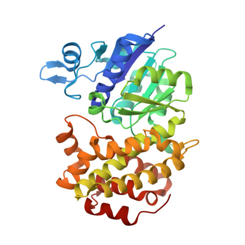
    Homo sapiens L-alpha-glycerol-3-phosphate dehydrogenase 1 (GPD1) catalyzes the reversible biological conversion of dihydroxyacetone (DHAP) to glycerol-3-phosphate. The GPD1 protein was expressed in Escherichia coli, and purified as a fusion protein with glutathione S-transferase. Here we report the apoenzyme structure of GPD1 determined by multiwavelength anomalous diffraction phasing, and other complex structures with small molecules (NAD+ and DHAP) by the molecular replacement method. This enzyme structure is organized into two distinct domains, the N-terminal eight-stranded beta-sheet sandwich domain and the C-terminal helical substrate-binding domain. An electrophilic catalytic mechanism by the epsilon-NH3+ group of Lys204 is proposed on the basis of the structural analyses. In addition, the inhibitory effects of zinc and sulfate on GPDHs are assayed and discussed.


  • Organizational Affiliation

    National Laboratory of Biomacromolecules, Institute of Biophysics (IBP), Chinese Academy of Sciences, Beijing 100101, China.


Macromolecules
Find similar proteins by:  (by identity cutoff)  |  3D Structure
Entity ID: 1
MoleculeChains Sequence LengthOrganismDetailsImage
Glycerol-3-phosphate dehydrogenase [NAD+], cytoplasmic
A, B
349Homo sapiensMutation(s): 0 
EC: 1.1.1.8
UniProt & NIH Common Fund Data Resources
Find proteins for P21695 (Homo sapiens)
Explore P21695 
Go to UniProtKB:  P21695
PHAROS:  P21695
GTEx:  ENSG00000167588 
Entity Groups  
Sequence Clusters30% Identity50% Identity70% Identity90% Identity95% Identity100% Identity
UniProt GroupP21695
Sequence Annotations
Expand
  • Reference Sequence
Small Molecules
Ligands 3 Unique
IDChains Name / Formula / InChI Key2D Diagram3D Interactions
NAD
Query on NAD

Download Ideal Coordinates CCD File 
J [auth A],
M [auth B]
NICOTINAMIDE-ADENINE-DINUCLEOTIDE
C21 H27 N7 O14 P2
BAWFJGJZGIEFAR-NNYOXOHSSA-N
13P
Query on 13P

Download Ideal Coordinates CCD File 
I [auth A],
L [auth B]
1,3-DIHYDROXYACETONEPHOSPHATE
C3 H7 O6 P
GNGACRATGGDKBX-UHFFFAOYSA-N
SO4
Query on SO4

Download Ideal Coordinates CCD File 
C [auth A]
D [auth A]
E [auth A]
F [auth A]
G [auth A]
C [auth A],
D [auth A],
E [auth A],
F [auth A],
G [auth A],
H [auth A],
K [auth B]
SULFATE ION
O4 S
QAOWNCQODCNURD-UHFFFAOYSA-L
Experimental Data & Validation

Experimental Data

  • Method: X-RAY DIFFRACTION
  • Resolution: 2.50 Å
  • R-Value Free: 0.294 
  • R-Value Work: 0.258 
  • R-Value Observed: 0.263 
  • Space Group: P 43 21 2
Unit Cell:
Length ( Å )Angle ( ˚ )
a = 115.995α = 90
b = 115.995β = 90
c = 153.72γ = 90
Software Package:
Software NamePurpose
DENZOdata reduction
SCALEPACKdata scaling
CNSrefinement
CNSphasing

Structure Validation

View Full Validation Report


Ligand Structure Quality Assessment 


Entry History 

Deposition Data

Revision History  (Full details and data files)

  • Version 1.0: 2006-04-11
    Type: Initial release
  • Version 1.1: 2008-04-30
    Changes: Version format compliance
  • Version 1.2: 2011-07-13
    Changes: Version format compliance
  • Version 1.3: 2024-03-13
    Changes: Data collection, Database references, Derived calculations