1WAA

IG27 protein domain


Experimental Data Snapshot

  • Method: X-RAY DIFFRACTION
  • Resolution: 1.80 Å
  • R-Value Free: 0.268 
  • R-Value Work: 0.207 
  • R-Value Observed: 0.211 

wwPDB Validation   3D Report Full Report


This is version 1.3 of the entry. See complete history


Literature

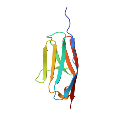
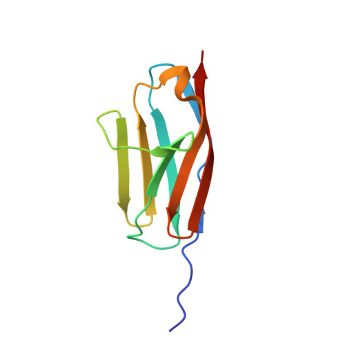
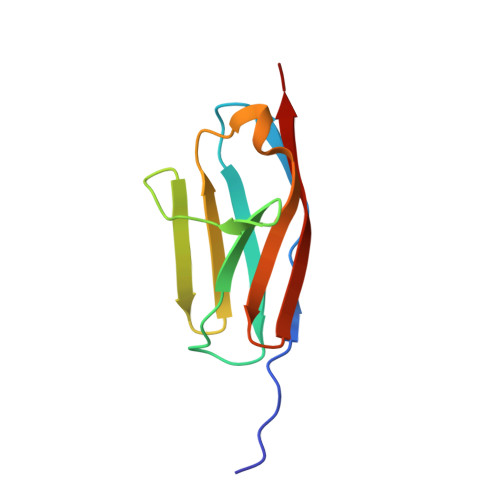
Mechanical Network in Titin Immunoglobulin from Force Distribution Analysis.

Stacklies, W.Vega, M.C.Wilmanns, M.Grater, F.

(2009) PLoS Comput Biol 5: 00306

  • DOI: https://doi.org/10.1371/journal.pcbi.1000306
  • Primary Citation of Related Structures:  
    1WAA

  • PubMed Abstract: 

    The role of mechanical force in cellular processes is increasingly revealed by single molecule experiments and simulations of force-induced transitions in proteins. How the applied force propagates within proteins determines their mechanical behavior yet remains largely unknown. We present a new method based on molecular dynamics simulations to disclose the distribution of strain in protein structures, here for the newly determined high-resolution crystal structure of I27, a titin immunoglobulin (IG) domain. We obtain a sparse, spatially connected, and highly anisotropic mechanical network. This allows us to detect load-bearing motifs composed of interstrand hydrogen bonds and hydrophobic core interactions, including parts distal to the site to which force was applied. The role of the force distribution pattern for mechanical stability is tested by in silico unfolding of I27 mutants. We then compare the observed force pattern to the sparse network of coevolved residues found in this family. We find a remarkable overlap, suggesting the force distribution to reflect constraints for the evolutionary design of mechanical resistance in the IG family. The force distribution analysis provides a molecular interpretation of coevolution and opens the road to the study of the mechanism of signal propagation in proteins in general.


  • Organizational Affiliation

    CAS-MPG Partner Institute for Computational Biology, Shanghai, People's Republic of China.


Macromolecules
Find similar proteins by:  (by identity cutoff)  |  3D Structure
Entity ID: 1
MoleculeChains Sequence LengthOrganismDetailsImage
TITIN
A, B, C, D
93Homo sapiensMutation(s): 0 
EC: 2.7.1
UniProt & NIH Common Fund Data Resources
Find proteins for Q8WZ42 (Homo sapiens)
Explore Q8WZ42 
Go to UniProtKB:  Q8WZ42
PHAROS:  Q8WZ42
GTEx:  ENSG00000155657 
Entity Groups  
Sequence Clusters30% Identity50% Identity70% Identity90% Identity95% Identity100% Identity
UniProt GroupQ8WZ42
Sequence Annotations
Expand
  • Reference Sequence
Find similar proteins by:  (by identity cutoff)  |  3D Structure
Entity ID: 2
MoleculeChains Sequence LengthOrganismDetailsImage
TITIN93Homo sapiensMutation(s): 0 
EC: 2.7.1
UniProt & NIH Common Fund Data Resources
Find proteins for Q8WZ42 (Homo sapiens)
Explore Q8WZ42 
Go to UniProtKB:  Q8WZ42
PHAROS:  Q8WZ42
GTEx:  ENSG00000155657 
Entity Groups  
Sequence Clusters30% Identity50% Identity70% Identity90% Identity95% Identity100% Identity
UniProt GroupQ8WZ42
Sequence Annotations
Expand
  • Reference Sequence
Find similar proteins by:  (by identity cutoff)  |  3D Structure
Entity ID: 3
MoleculeChains Sequence LengthOrganismDetailsImage
TITIN93Homo sapiensMutation(s): 0 
EC: 2.7.1
UniProt & NIH Common Fund Data Resources
Find proteins for Q8WZ42 (Homo sapiens)
Explore Q8WZ42 
Go to UniProtKB:  Q8WZ42
PHAROS:  Q8WZ42
GTEx:  ENSG00000155657 
Entity Groups  
Sequence Clusters30% Identity50% Identity70% Identity90% Identity95% Identity100% Identity
UniProt GroupQ8WZ42
Sequence Annotations
Expand
  • Reference Sequence
Small Molecules
Ligands 1 Unique
IDChains Name / Formula / InChI Key2D Diagram3D Interactions
ZN
Query on ZN

Download Ideal Coordinates CCD File 
AA [auth E]
BA [auth F]
CA [auth F]
DA [auth F]
G [auth A]
AA [auth E],
BA [auth F],
CA [auth F],
DA [auth F],
G [auth A],
H [auth A],
I [auth A],
J [auth A],
K [auth A],
L [auth B],
M [auth B],
N [auth B],
O [auth B],
P [auth B],
Q [auth C],
R [auth C],
S [auth C],
T [auth C],
U [auth C],
V [auth D],
W [auth D],
X [auth D],
Y [auth E],
Z [auth E]
ZINC ION
Zn
PTFCDOFLOPIGGS-UHFFFAOYSA-N
Experimental Data & Validation

Experimental Data

  • Method: X-RAY DIFFRACTION
  • Resolution: 1.80 Å
  • R-Value Free: 0.268 
  • R-Value Work: 0.207 
  • R-Value Observed: 0.211 
  • Space Group: P 21 21 21
Unit Cell:
Length ( Å )Angle ( ˚ )
a = 62.24α = 90
b = 75.99β = 90
c = 134.22γ = 90
Software Package:
Software NamePurpose
REFMACrefinement
MOSFLMdata reduction
SCALAdata scaling
AMoREphasing

Structure Validation

View Full Validation Report


Entry History 

Deposition Data

Revision History  (Full details and data files)

  • Version 1.0: 2006-07-05
    Type: Initial release
  • Version 1.1: 2011-12-28
    Changes: Database references, Derived calculations, Non-polymer description, Other, Refinement description, Version format compliance
  • Version 1.2: 2019-07-24
    Changes: Advisory, Data collection
  • Version 1.3: 2023-12-13
    Changes: Advisory, Data collection, Database references, Derived calculations, Refinement description