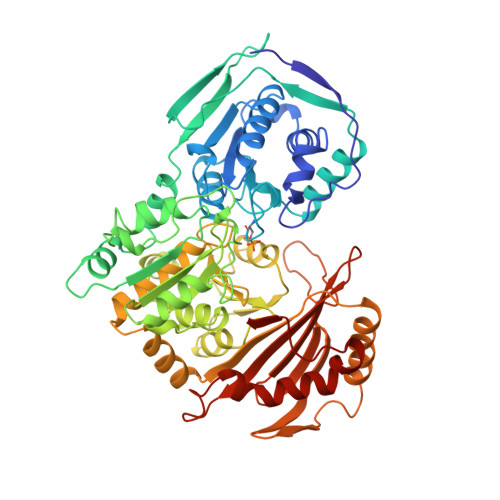
Structural changes at the metal ion binding site during the phosphoglucomutase reaction.
Ray Jr., W.J., Post, C.B., Liu, Y., Rhyu, G.I.(1993) Biochemistry 32: 48-57
- PubMed: 8418859
- DOI: https://doi.org/10.1021/bi00052a008
- Primary Citation of Related Structures:
1VKL - PubMed Abstract:
An electron density map of the reactive, Cd2+ form of crystalline phosphoglucomutase from X-ray diffraction studies shows that the enzymic phosphate donates a nonbridging oxygen to the ligand sphere of the bound metal ion, which appears to be tetracoordinate. 31P and 113Cd NMR spectroscopy are used to assess changes in the properties of bound Cd2+ produced by substrate/product and by substrate/product analog inhibitors. The approximately 50 ppm downfield shift of the 113Cd resonance on formation of the complex of dephosphoenzyme and glucose 1,6-bisphosphate is associated with the initial sugar-phosphate binding step and likely involves a change in the geometry of the coordinating ligands. This interpretation is supported by spectral studies involving various complexes of the active Co2+ and Ni(2+)-enzyme. In addition, there is a loss of the 31P-113Cd J coupling that characterizes the monophosphate complexes of the Cd2+ enzyme either during or immediately after the PO3- transfer step that produces the bisphosphate complex, indicating a further change at the metal binding site. The implications of these observations with respect to the PO3- transfer process in the phosphoglucomutase reaction are considered. The apparent plasticity of the ligand sphere of the active site metal ion in this system may allow a single metal ion to act as a chaperone for a nonbridging oxygen during PO3- transfer or to allow a change in metal ion coordination during catalysis. A general NMR line shape/chemical-exchange analysis for evaluating binding in protein-ligand systems when exchange is intermediate to fast on the NMR time scale is described. Its application to the present system involves multiple exchange sites that depend on a single binding rate, thereby adding further constraints to the analysis.
Organizational Affiliation:
Department of Biological Sciences, Purdue University, West Lafayette, Indiana 47907.