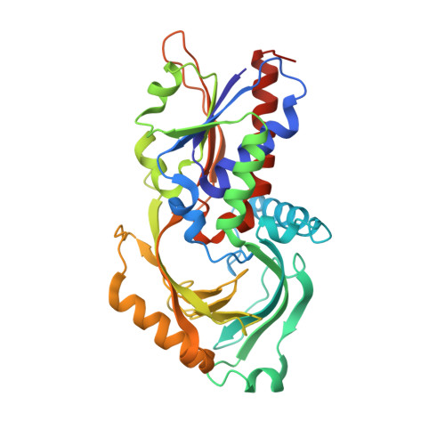
1VE9

Porcine kidney D-amino acid oxidase


Experimental Data Snapshot

  • Method: X-RAY DIFFRACTION
  • Resolution: 2.50 Å
  • R-Value Free: 0.254 
  • R-Value Work: 0.187 

wwPDB Validation   3D Report Full Report


Ligand Structure Quality Assessment 


This is version 1.5 of the entry. See complete history


Literature

Three-dimensional structure of porcine kidney D-amino acid oxidase at 3.0 A resolution.

Mizutani, H.Miyahara, I.Hirotsu, K.Nishina, Y.Shiga, K.Setoyama, C.Miura, R.

(1996) J Biochem 120: 14-17

  • DOI: https://doi.org/10.1093/oxfordjournals.jbchem.a021376
  • Primary Citation of Related Structures:  
    1VE9

  • PubMed Abstract: 

    The X-ray crystallographic structure of porcine kidney D-amino acid oxidase, which had been expressed in Escherichia coli transformed with a vector containing DAO cDNA, was determined by the isomorphous replacement method for the complex form with benzoate. The known amino acid sequence, FAD and benzoate were fitted to an electron density map of 3.0 A resolution with an R-factor of 21.0%. The overall dimeric structure exhibits an elongated ellipsoidal framework. The prosthetic group, FAD, was found to be in an extended conformation, the isoalloxazine ring being buried in the protein core. The ADP moiety of FAD was located in the typical beta alpha beta dinucleotide binding motif, with the alpha-helix dipole stabilizing the pyrophosphate negative charge. The substrate analog, benzoate, is located on the re-face of the isoalloxazine ring, while the si-face is blocked by hydrophobic residues. The carboxylate group of benzoate is ion-paired with the Arg283 side chain and is within interacting distance with the hydroxy moiety of Tyr228. The phenol ring of Tyr224 is located just above the benzene ring of benzoate, implying the importance of this residue for catalysis. There is no positive charge or alpha-helix dipole near N(1) of flavin. Hydrogen bonds were observed at C(2) = O, N(3)-H, C(4) = O, and N(5) of the flavin ring.


  • Organizational Affiliation

    Department of Chemistry, Faculty of Science, Osaka City University.


Macromolecules
Find similar proteins by:  (by identity cutoff)  |  3D Structure
Entity ID: 1
MoleculeChains Sequence LengthOrganismDetailsImage
D-amino acid oxidase
A, B
347Sus scrofaMutation(s): 0 
EC: 1.4.3.3
UniProt
Find proteins for P00371 (Sus scrofa)
Explore P00371 
Go to UniProtKB:  P00371
Entity Groups  
Sequence Clusters30% Identity50% Identity70% Identity90% Identity95% Identity100% Identity
UniProt GroupP00371
Sequence Annotations
Expand
  • Reference Sequence
Small Molecules
Binding Affinity Annotations 
IDSourceBinding Affinity
BEZ BindingDB:  1VE9 IC50: 1.34e+5 (nM) from 1 assay(s)
Experimental Data & Validation

Experimental Data

  • Method: X-RAY DIFFRACTION
  • Resolution: 2.50 Å
  • R-Value Free: 0.254 
  • R-Value Work: 0.187 
  • Space Group: P 21 21 21
Unit Cell:
Length ( Å )Angle ( ˚ )
a = 110.3α = 90
b = 92.9β = 90
c = 71.6γ = 90
Software Package:
Software NamePurpose
CCP4model building
CCP4phasing
CNSrefinement

Structure Validation

View Full Validation Report


Ligand Structure Quality Assessment 


Entry History 

Deposition Data

Revision History  (Full details and data files)

  • Version 1.0: 2004-04-13
    Type: Initial release
  • Version 1.1: 2008-04-27
    Changes: Version format compliance
  • Version 1.2: 2011-07-13
    Changes: Version format compliance
  • Version 1.3: 2017-10-04
    Changes: Refinement description
  • Version 1.4: 2019-11-06
    Changes: Data collection, Database references
  • Version 1.5: 2023-12-27
    Changes: Data collection, Database references, Derived calculations