1VCM

Crystal Structure of T.th. HB8 CTP synthetase


Experimental Data Snapshot

  • Method: X-RAY DIFFRACTION
  • Resolution: 2.35 Å
  • R-Value Free: 0.279 
  • R-Value Work: 0.235 

wwPDB Validation   3D Report Full Report


This is version 1.3 of the entry. See complete history


Literature

Crystal Structures of CTP Synthetase Reveal ATP, UTP, and Glutamine Binding Sites

Goto, M.Omi, R.Nakagawa, N.Miyahara, I.Hirotsu, K.

(2004) Structure 12: 1413-1423

  • DOI: https://doi.org/10.1016/j.str.2004.05.013
  • Primary Citation of Related Structures:  
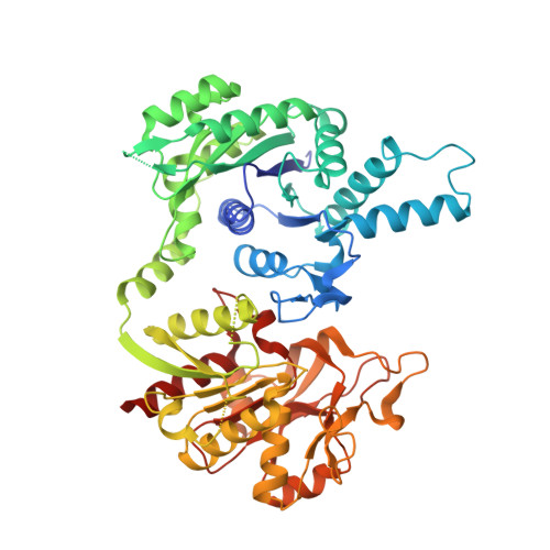
    1VCM, 1VCN, 1VCO

  • PubMed Abstract: 

    CTP synthetase (CTPs) catalyzes the last step in CTP biosynthesis, in which ammonia generated at the glutaminase domain reacts with the ATP-phosphorylated UTP at the synthetase domain to give CTP. Glutamine hydrolysis is active in the presence of ATP and UTP and is stimulated by the addition of GTP. We report the crystal structures of Thermus thermophilus HB8 CTPs alone, CTPs with 3SO4(2-), and CTPs with glutamine. The enzyme is folded into a homotetramer with a cross-shaped structure. Based on the binding mode of sulfate anions to the synthetase site, ATP and UTP are computer modeled into CTPs with a geometry favorable for the reaction. Glutamine bound to the glutaminase domain is situated next to the triad of Glu-His-Cys as a catalyst and a water molecule. Structural information provides an insight into the conformational changes associated with the binding of ATP and UTP and the formation of the GTP binding site.


  • Organizational Affiliation

    Department of Chemistry, Graduate School of Science, Osaka University, Osaka 560-0043, Japan.


Macromolecules
Find similar proteins by:  (by identity cutoff)  |  3D Structure
Entity ID: 1
MoleculeChains Sequence LengthOrganismDetailsImage
CTP synthetase550Thermus thermophilusMutation(s): 0 
Gene Names: HB8
EC: 6.3.4.2
UniProt
Find proteins for Q5SIA8 (Thermus thermophilus (strain ATCC 27634 / DSM 579 / HB8))
Explore Q5SIA8 
Go to UniProtKB:  Q5SIA8
Entity Groups  
Sequence Clusters30% Identity50% Identity70% Identity90% Identity95% Identity100% Identity
UniProt GroupQ5SIA8
Sequence Annotations
Expand
  • Reference Sequence
Experimental Data & Validation

Experimental Data

  • Method: X-RAY DIFFRACTION
  • Resolution: 2.35 Å
  • R-Value Free: 0.279 
  • R-Value Work: 0.235 
  • Space Group: I 2 2 2
Unit Cell:
Length ( Å )Angle ( ˚ )
a = 86α = 90
b = 120.67β = 90
c = 143.13γ = 90
Software Package:
Software NamePurpose
HKL-2000data collection
SCALEPACKdata scaling
AMoREphasing
CNSrefinement
HKL-2000data reduction

Structure Validation

View Full Validation Report


Entry History 

Deposition Data

Revision History  (Full details and data files)

  • Version 1.0: 2004-08-31
    Type: Initial release
  • Version 1.1: 2008-04-27
    Changes: Version format compliance
  • Version 1.2: 2011-07-13
    Changes: Derived calculations, Version format compliance
  • Version 1.3: 2023-12-27
    Changes: Data collection, Database references