1V0H

ASCOBATE PEROXIDASE FROM SOYBEAN CYTOSOL IN COMPLEX WITH SALICYLHYDROXAMIC ACID


Experimental Data Snapshot

  • Method: X-RAY DIFFRACTION
  • Resolution: 1.46 Å
  • R-Value Free: 0.182 
  • R-Value Work: 0.150 
  • R-Value Observed: 0.151 

wwPDB Validation   3D Report Full Report


Ligand Structure Quality Assessment 


This is version 1.4 of the entry. See complete history


Literature

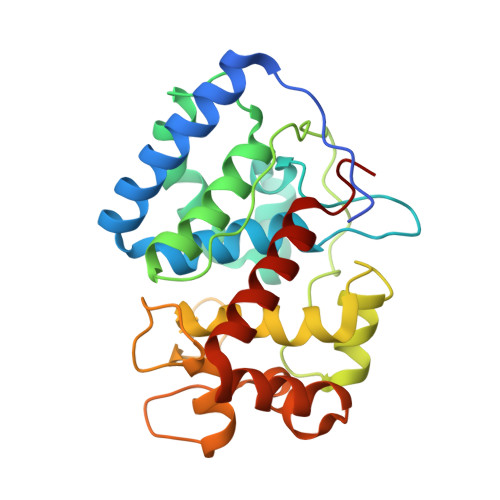
Crystal Structure of the Ascorbate Peroxidase-Salicylhydroxamic Acid Complex

Sharp, K.H.Moody, P.C.E.Brown, K.A.Raven, E.L.

(2004) Biochemistry 43: 8644

  • DOI: https://doi.org/10.1021/bi049343q
  • Primary Citation of Related Structures:  
    1V0H

  • PubMed Abstract: 

    Ascorbate peroxidase is a bifunctional peroxidase that catalyzes the H(2)O(2)-dependent oxidation of both ascorbate and various aromatic substrates. The ascorbate binding site was recently identified as being close to the gamma-heme edge [Sharp, K. H., Mewies, M., Moody, P. C. E., and Raven, E. L. (2003)Nat. Struct. Biol. 10, 303-307]. In this work, the X-ray crystal structure of recombinant soybean cytosolic ascorbate peroxidase (rsAPX) in complex with salicylhydroxamic acid (SHA) has been determined to 1.46 A. The SHA molecule is bound close to the delta-heme edge in a cavity that connects the distal side of the heme to the surface of the protein. There are hydrogen bonds between the phenolic hydroxide of the SHA and the main chain carbonyl of Pro132, between the carbonyl oxygen of SHA and the side chain guanadinium group of Arg38, and between the hydroxamic acid group and the indole nitrogen of Trp41. The structure provides the first information about the location of the aromatic binding site in ascorbate peroxidase and, together with our previous data [Sharp, K. H., et al. (2003) Nat. Struct. Biol. 10, 303-307], completes the structural description of the binding properties of ascorbate peroxidase. The mechanistic implications of the results are discussed in terms of our current understanding of how APX catalyzes oxidation of different types of substrates bound at different locations.


  • Organizational Affiliation

    Department of Chemistry, University of Leicester, UK.


Macromolecules
Find similar proteins by:  (by identity cutoff)  |  3D Structure
Entity ID: 1
MoleculeChains Sequence LengthOrganismDetailsImage
ASCORBATE PEROXIDASEA [auth X]261Glycine maxMutation(s): 0 
EC: 1.11.1.11
UniProt
Find proteins for Q43758 (Glycine max)
Explore Q43758 
Go to UniProtKB:  Q43758
Entity Groups  
Sequence Clusters30% Identity50% Identity70% Identity90% Identity95% Identity100% Identity
UniProt GroupQ43758
Sequence Annotations
Expand
  • Reference Sequence
Experimental Data & Validation

Experimental Data

  • Method: X-RAY DIFFRACTION
  • Resolution: 1.46 Å
  • R-Value Free: 0.182 
  • R-Value Work: 0.150 
  • R-Value Observed: 0.151 
  • Space Group: P 42 21 2
Unit Cell:
Length ( Å )Angle ( ˚ )
a = 82.77α = 90
b = 82.77β = 90
c = 74.991γ = 90
Software Package:
Software NamePurpose
REFMACrefinement
DENZOdata reduction
SCALEPACKdata scaling

Structure Validation

View Full Validation Report


Ligand Structure Quality Assessment 


Entry History 

Deposition Data

Revision History  (Full details and data files)

  • Version 1.0: 2004-06-23
    Type: Initial release
  • Version 1.1: 2011-05-07
    Changes: Version format compliance
  • Version 1.2: 2011-07-13
    Changes: Version format compliance
  • Version 1.3: 2018-10-24
    Changes: Data collection, Derived calculations, Source and taxonomy
  • Version 1.4: 2023-12-13
    Changes: Data collection, Database references, Derived calculations, Other, Refinement description