


The Crystal Structure of Molybdopterin Biosynthesis Moeaprotein from Pyrococcus Horikosii
Takahashi, H., Miyano, M., Tahirov, T.H.To be published.
Experimental Data Snapshot
wwPDB Validation 3D Report Full Report

Entity ID: 1 | |||||
|---|---|---|---|---|---|
| Molecule | Chains | Sequence Length | Organism | Details | Image |
| 402AA LONG HYPOTHETICAL MOLYBDOPTERIN BIOSYNTHESIS MOEA PROTEIN | 402 | Pyrococcus horikoshii OT3 | Mutation(s): 0 | 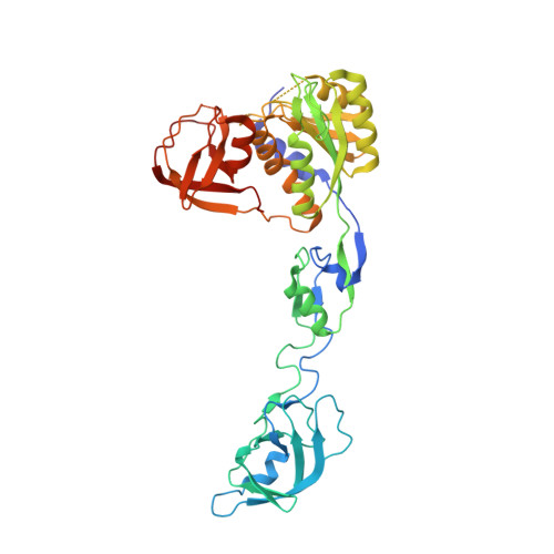 | |
UniProt | |||||
Find proteins for O58335 (Pyrococcus horikoshii (strain ATCC 700860 / DSM 12428 / JCM 9974 / NBRC 100139 / OT-3)) Explore O58335 Go to UniProtKB: O58335 | |||||
Entity Groups | |||||
| Sequence Clusters | 30% Identity50% Identity70% Identity90% Identity95% Identity100% Identity | ||||
| UniProt Group | O58335 | ||||
Sequence AnnotationsExpand | |||||
| |||||
| Ligands 1 Unique | |||||
|---|---|---|---|---|---|
| ID | Chains | Name / Formula / InChI Key | 2D Diagram | 3D Interactions | |
| SO4 Query on SO4 | B [auth A] C [auth A] D [auth A] E [auth A] F [auth A] | SULFATE ION O4 S QAOWNCQODCNURD-UHFFFAOYSA-L |  | ||
| Length ( Å ) | Angle ( ˚ ) |
|---|---|
| a = 181.375 | α = 90 |
| b = 181.375 | β = 90 |
| c = 71.311 | γ = 120 |
| Software Name | Purpose |
|---|---|
| CNS | refinement |
| DENZO | data reduction |
| SCALEPACK | data scaling |
| SOLVE | phasing |
RCSB PDB (citation) is hosted by
RCSB PDB is a member of the

