1UMN

Crystal structure of Dps-like peroxide resistance protein (Dpr) from Streptococcus suis


Experimental Data Snapshot

  • Method: X-RAY DIFFRACTION
  • Resolution: 1.95 Å
  • R-Value Free: 0.224 
  • R-Value Work: 0.185 
  • R-Value Observed: 0.185 

wwPDB Validation   3D Report Full Report


This is version 1.4 of the entry. See complete history


Literature

Crystal structure of Streptococcus suis Dps-like peroxide resistance protein Dpr: implications for iron incorporation.

Kauko, A.Haataja, S.Pulliainen, A.T.Finne, J.Papageorgiou, A.C.

(2004) J Mol Biol 338: 547-558

  • DOI: https://doi.org/10.1016/j.jmb.2004.03.009
  • Primary Citation of Related Structures:  
    1UMN

  • PubMed Abstract: 

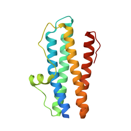
    The Dps-like peroxide resistance protein (Dpr) is an aerotolerance and hydrogen peroxide resistance agent found in the meningitis-associated pathogen Streptococcus suis. Dpr is believed to act by binding free intracellular iron to prevent Fenton chemistry-catalysed formation of toxic hydroxyl radicals. The crystal structure of Dpr has been determined to 1.95 A resolution. The final model has an Rcyst value of 18.5% (Rfree = 22.4%) and consists of 12 identical monomers (each of them comprising a four alpha-helix bundle) that form a hollow sphere obeying 23 symmetry. Structural features show that Dpr belongs to the Dps family of bacterial proteins. Twelve putative ferroxidase centers, each formed at the interface of neighboring monomer pairs, were identified in the Dpr structure with structural similarities to those found in other Dps family members. Dpr was crystallized in the absence of iron, hence no bound iron was found in the structure in contrast to other Dps family members. A novel metal-binding site approximately 6A from the ferroxidase centre was identified and assigned to a bound calcium ion. Two residues from the ferroxidase centre (Asp63 and Asp74) were found to be involved in calcium binding. Structural comparison with other family members revealed that Asp63 and Asp74 adopt different conformation in the Dpr structure. The structure of Dpr presented here shows potential local conformational changes that may occur during iron incorporation. A role for the metal-binding site in iron uptake is proposed.


  • Organizational Affiliation

    Turku Centre for Biotechnology, University of Turku and Abo Akademi University, BioCity, Turku 20521, Finland.


Macromolecules
Find similar proteins by:  (by identity cutoff)  |  3D Structure
Entity ID: 1
MoleculeChains Sequence LengthOrganismDetailsImage
DPS-LIKE PEROXIDE RESISTANCE PROTEIN
A, B, C, D, E
A, B, C, D, E, F, G, H, I, J, K, L
165Streptococcus suisMutation(s): 0 
UniProt
Find proteins for P0CB53 (Streptococcus suis)
Explore P0CB53 
Go to UniProtKB:  P0CB53
Entity Groups  
Sequence Clusters30% Identity50% Identity70% Identity90% Identity95% Identity100% Identity
UniProt GroupP0CB53
Sequence Annotations
Expand
  • Reference Sequence
Small Molecules
Ligands 3 Unique
IDChains Name / Formula / InChI Key2D Diagram3D Interactions
EPE
Query on EPE

Download Ideal Coordinates CCD File 
DA [auth J],
FA [auth K],
S [auth E],
Z [auth I]
4-(2-HYDROXYETHYL)-1-PIPERAZINE ETHANESULFONIC ACID
C8 H18 N2 O4 S
JKMHFZQWWAIEOD-UHFFFAOYSA-N
CA
Query on CA

Download Ideal Coordinates CCD File 
BA [auth I]
CA [auth I]
EA [auth J]
GA [auth K]
HA [auth K]
BA [auth I],
CA [auth I],
EA [auth J],
GA [auth K],
HA [auth K],
IA [auth L],
M [auth A],
N [auth B],
O [auth C],
Q [auth D],
R [auth D],
T [auth E],
U [auth F],
V [auth G],
W [auth G],
Y [auth H]
CALCIUM ION
Ca
BHPQYMZQTOCNFJ-UHFFFAOYSA-N
CL
Query on CL

Download Ideal Coordinates CCD File 
AA [auth I],
P [auth D],
X [auth H]
CHLORIDE ION
Cl
VEXZGXHMUGYJMC-UHFFFAOYSA-M
Experimental Data & Validation

Experimental Data

  • Method: X-RAY DIFFRACTION
  • Resolution: 1.95 Å
  • R-Value Free: 0.224 
  • R-Value Work: 0.185 
  • R-Value Observed: 0.185 
  • Space Group: P 21 21 21
Unit Cell:
Length ( Å )Angle ( ˚ )
a = 104.735α = 90
b = 138.168β = 90
c = 142.707γ = 90
Software Package:
Software NamePurpose
CNSrefinement
DENZOdata reduction
SCALEPACKdata scaling
AMoREphasing

Structure Validation

View Full Validation Report


Entry History 

Deposition Data

Revision History  (Full details and data files)

  • Version 1.0: 2004-04-23
    Type: Initial release
  • Version 1.1: 2011-05-08
    Changes: Version format compliance
  • Version 1.2: 2011-07-13
    Changes: Version format compliance
  • Version 1.3: 2019-03-13
    Changes: Data collection, Database references, Experimental preparation, Other
  • Version 1.4: 2023-12-13
    Changes: Data collection, Database references, Derived calculations, Other, Refinement description