1UKM

Crystal structure of EMS16, an Antagonist of collagen receptor integrin alpha2beta1 (GPIa/IIa)


Experimental Data Snapshot

  • Method: X-RAY DIFFRACTION
  • Resolution: 1.90 Å
  • R-Value Free: 0.233 
  • R-Value Work: 0.196 
  • R-Value Observed: 0.196 

wwPDB Validation   3D Report Full Report


Ligand Structure Quality Assessment 


This is version 1.6 of the entry. See complete history


Literature

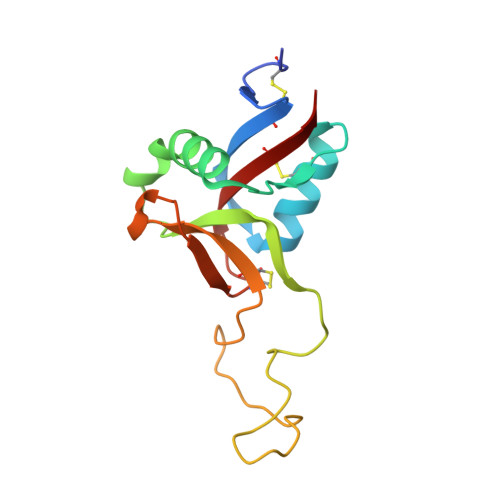
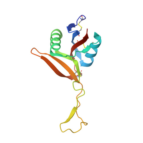
Structural characterization of EMS16, an Antagonist of collagen receptor (GPIa/IIa) from the venom of Echis multisquamatus

Horii, K.Okuda, D.Morita, T.Mizuno, H.

(2003) Biochemistry 42: 12497-12502

  • DOI: https://doi.org/10.1021/bi034890h
  • Primary Citation of Related Structures:  
    1UKM

  • PubMed Abstract: 

    Snake venoms contain a number of hemostatically active C-type lectin-like proteins (CLPs), which affect the blood coagulation system, endothelial cells, and platelets. CLPs have broad similarities in structure and possess distinct biological functions. EMS16, a CLP from Echis multisquamatus venom, which is a potent and selective inhibitor of the collagen receptor, glycoprotein Ia/IIa (integrin alpha2beta1), has been used in the present study to examine structure-function relationships in venom CLPs by X-ray crystallography. The structure of EMS16, determined at a resolution of 1.9 A, revealed a heterodimer involved with domain swapping of the central loop as observed in the structures of other CLPs. A part of the glycan was observed and identified as N-acetyl-D-glucosamine (GlcNAc) in the electron density map at Asn21 of subunit B, an expected glycosylation site. EMS16 had a unique, positively charged electrostatic potential patch on the concave surface that may qualify as a site for interaction with the I-domain of the glycoprotein Ia/IIa.


  • Organizational Affiliation

    Department of Biochemistry, National Institute of Agrobiological Sciences, 2-1-2 Kannondai, Tsukuba, Ibaraki 305-8602, Japan.


Macromolecules
Find similar proteins by:  (by identity cutoff)  |  3D Structure
Entity ID: 1
MoleculeChains Sequence LengthOrganismDetailsImage
EMS16 A chain134Echis multisquamatusMutation(s): 0 
UniProt
Find proteins for Q7T2Q1 (Echis multisquamatus)
Explore Q7T2Q1 
Go to UniProtKB:  Q7T2Q1
Entity Groups  
Sequence Clusters30% Identity50% Identity70% Identity90% Identity95% Identity100% Identity
UniProt GroupQ7T2Q1
Sequence Annotations
Expand
  • Reference Sequence
Find similar proteins by:  (by identity cutoff)  |  3D Structure
Entity ID: 2
MoleculeChains Sequence LengthOrganismDetailsImage
EMS16 B chain128Echis multisquamatusMutation(s): 0 
UniProt
Find proteins for Q7T2Q0 (Echis multisquamatus)
Explore Q7T2Q0 
Go to UniProtKB:  Q7T2Q0
Entity Groups  
Sequence Clusters30% Identity50% Identity70% Identity90% Identity95% Identity100% Identity
UniProt GroupQ7T2Q0
Sequence Annotations
Expand
  • Reference Sequence
Experimental Data & Validation

Experimental Data

  • Method: X-RAY DIFFRACTION
  • Resolution: 1.90 Å
  • R-Value Free: 0.233 
  • R-Value Work: 0.196 
  • R-Value Observed: 0.196 
  • Space Group: P 21 21 21
Unit Cell:
Length ( Å )Angle ( ˚ )
a = 46.57α = 90
b = 59.933β = 90
c = 115.743γ = 90
Software Package:
Software NamePurpose
CNSrefinement
AMoREphasing

Structure Validation

View Full Validation Report


Ligand Structure Quality Assessment 


Entry History 

Deposition Data

Revision History  (Full details and data files)

  • Version 1.0: 2003-11-04
    Type: Initial release
  • Version 1.1: 2008-04-27
    Changes: Version format compliance
  • Version 1.2: 2011-07-13
    Changes: Non-polymer description, Version format compliance
  • Version 1.3: 2017-10-04
    Changes: Refinement description
  • Version 1.4: 2018-07-25
    Changes: Data collection, Database references, Structure summary
  • Version 1.5: 2020-07-29
    Type: Remediation
    Reason: Carbohydrate remediation
    Changes: Data collection, Derived calculations, Structure summary
  • Version 1.6: 2023-10-25
    Changes: Data collection, Database references, Refinement description, Structure summary