1TZL

Crystal Structure of Pyranose 2-Oxidase from the White-Rot Fungus Peniophora sp.


Experimental Data Snapshot

  • Method: X-RAY DIFFRACTION
  • Resolution: 2.35 Å
  • R-Value Free: 0.226 
  • R-Value Work: 0.174 
  • R-Value Observed: 0.176 

wwPDB Validation   3D Report Full Report


Ligand Structure Quality Assessment 


This is version 1.3 of the entry. See complete history


Literature

Crystal structure of pyranose 2-oxidase from the white-rot fungus peniophora sp.

Bannwarth, M.Bastian, S.Heckmann-Pohl, D.Giffhorn, F.Schulz, G.E.

(2004) Biochemistry 43: 11683-11690

  • DOI: https://doi.org/10.1021/bi048609q
  • Primary Citation of Related Structures:  
    1TZL

  • PubMed Abstract: 

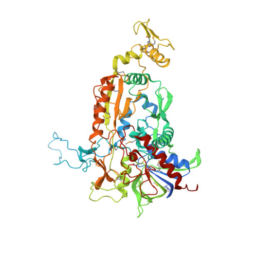
    Pyranose 2-oxidase catalyzes the oxidation of a number of carbohydrates using dioxygen. The enzyme forms a D(2) symmetric homotetramer and contains one covalently bound FAD per subunit. The structure of the enzyme from Peniophora sp. was determined by multiwavelength anomalous diffraction (MAD) based on 96 selenium sites per crystallographic asymmetric unit and subsequently refined to good-quality indices. According to its chain fold, the enzyme belongs to the large glutathione reductase family and, in a more narrow sense, to the glucose-methanol-choline oxidoreductase (GMC) family. The tetramer contains a spacious central cavity from which the substrate enters one of the four active centers by penetrating a mobile barrier. Since this cavity can only be accessed by glucose-sized molecules, the enzyme does not convert sugars that are part of a larger molecule. The geometry of the active center and a comparison with an inhibitor complex of the homologous enzyme cellobiose dehydrogenase allow the modeling of the reaction at a high confidence level.


  • Organizational Affiliation

    Institut für Organische Chemie und Biochemie, Albert-Ludwigs-Universität, Albertstrasse 21, 79104 Freiburg im Breisgau, Germany.


Macromolecules
Find similar proteins by:  (by identity cutoff)  |  3D Structure
Entity ID: 1
MoleculeChains Sequence LengthOrganismDetailsImage
pyranose oxidase
A, B, C, D, E
A, B, C, D, E, F, G, H
622Peniophora sp. SGMutation(s): 16 
Gene Names: p2ox
EC: 1.1.3.10
UniProt
Find proteins for Q8J136 (Peniophora sp. (strain SG))
Explore Q8J136 
Go to UniProtKB:  Q8J136
Entity Groups  
Sequence Clusters30% Identity50% Identity70% Identity90% Identity95% Identity100% Identity
UniProt GroupQ8J136
Sequence Annotations
Expand
  • Reference Sequence
Experimental Data & Validation

Experimental Data

  • Method: X-RAY DIFFRACTION
  • Resolution: 2.35 Å
  • R-Value Free: 0.226 
  • R-Value Work: 0.174 
  • R-Value Observed: 0.176 
  • Space Group: P 1 21 1
Unit Cell:
Length ( Å )Angle ( ˚ )
a = 164.879α = 90
b = 103.596β = 105.25
c = 169.169γ = 90
Software Package:
Software NamePurpose
REFMACrefinement
MAR345data collection
XDSdata scaling
MLPHAREphasing

Structure Validation

View Full Validation Report


Ligand Structure Quality Assessment 


Entry History 

Deposition Data

Revision History  (Full details and data files)

  • Version 1.0: 2004-10-19
    Type: Initial release
  • Version 1.1: 2008-04-30
    Changes: Version format compliance
  • Version 1.2: 2011-07-13
    Changes: Version format compliance
  • Version 1.3: 2017-10-11
    Changes: Refinement description