1TJ7

Structure determination and refinement at 2.44 A resolution of Argininosuccinate lyase from E. coli


Experimental Data Snapshot

  • Method: X-RAY DIFFRACTION
  • Resolution: 2.44 Å
  • R-Value Free: 0.217 
  • R-Value Work: 0.167 
  • R-Value Observed: 0.169 

wwPDB Validation   3D Report Full Report


This is version 1.4 of the entry. See complete history


Literature

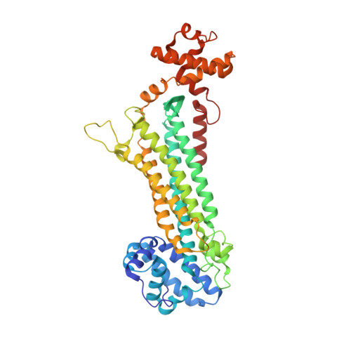
Structure determination and refinement at 2.44 A resolution of argininosuccinate lyase from Escherichia coli.

Bhaumik, P.Koski, M.K.Bergmann, U.Wierenga, R.K.

(2004) Acta Crystallogr D Biol Crystallogr 60: 1964-1970

  • DOI: https://doi.org/10.1107/S0907444904021912
  • Primary Citation of Related Structures:  
    1TJ7

  • PubMed Abstract: 

    Escherichia coli argininosuccinate lyase has been crystallized from a highly concentrated sample of purified recombinant alpha-methylacyl-CoA racemase, in which it occurred as a minor impurity. The structure has been solved using molecular replacement at 2.44 A resolution. The enzyme is tetrameric, but in this crystal form there is a dimer in the asymmetric unit. The tetramer has four active sites; each active site is constructed from loops of three different subunits. One of these catalytic loops, near residues Ser277 and Ser278, was disordered in previous structures of active lyases, but is very well ordered in this structure in one of the subunits owing to the presence of two phosphate ions in the active-site cavity. The positions of these phosphate ions indicate a plausible mode of binding of the succinate moiety of the substrate in the competent catalytic complex.


  • Organizational Affiliation

    Biocenter Oulu and Department of Biochemistry, University of Oulu, Linnanmaa, Box 3000, FIN-90014 Oulu, Finland.


Macromolecules
Find similar proteins by:  (by identity cutoff)  |  3D Structure
Entity ID: 1
MoleculeChains Sequence LengthOrganismDetailsImage
Argininosuccinate lyase
A, B
457Escherichia coliMutation(s): 0 
EC: 4.3.2.1
UniProt
Find proteins for P11447 (Escherichia coli (strain K12))
Explore P11447 
Go to UniProtKB:  P11447
Entity Groups  
Sequence Clusters30% Identity50% Identity70% Identity90% Identity95% Identity100% Identity
UniProt GroupP11447
Sequence Annotations
Expand
  • Reference Sequence
Experimental Data & Validation

Experimental Data

  • Method: X-RAY DIFFRACTION
  • Resolution: 2.44 Å
  • R-Value Free: 0.217 
  • R-Value Work: 0.167 
  • R-Value Observed: 0.169 
  • Space Group: P 61 2 2
Unit Cell:
Length ( Å )Angle ( ˚ )
a = 121.469α = 90
b = 121.469β = 90
c = 255.251γ = 120
Software Package:
Software NamePurpose
REFMACrefinement
MAR345data collection
SCALEPACKdata scaling
CNSphasing

Structure Validation

View Full Validation Report


Entry History 

Deposition Data

Revision History  (Full details and data files)

  • Version 1.0: 2004-10-26
    Type: Initial release
  • Version 1.1: 2008-04-30
    Changes: Version format compliance
  • Version 1.2: 2011-07-13
    Changes: Non-polymer description, Version format compliance
  • Version 1.3: 2017-10-11
    Changes: Data collection, Refinement description
  • Version 1.4: 2023-08-23
    Changes: Data collection, Database references, Derived calculations, Refinement description