1.42A crystal structure of mini-IGF-1(2): an analysis of the disulfide isomerization property and receptor binding property of IGF-1 based on the three-dimensional structure
Yun, C.H., Tang, Y.H., Feng, Y.M., An, X.M., Chang, W.R., Liang, D.C.(2004) Biochem Biophys Res Commun 326: 52-59
- PubMed: 15567151
- DOI: https://doi.org/10.1016/j.bbrc.2004.10.203
- Primary Citation of Related Structures:
1TGR - PubMed Abstract:
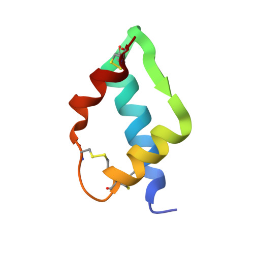
Insulin and insulin-like growth factor 1 (IGF-1) share a homologous sequence, a similar three-dimensional structure and weakly overlapping biological activity, but IGF-1 folds into two thermodynamically stable disulfide isomers, while insulin folds into one unique stable tertiary structure. This is a very interesting phenomenon in which one amino acid sequence encodes two three-dimensional structures, and its molecular mechanism has remained unclear for a long time. In this study, the crystal structure of mini-IGF-1(2), a disulfide isomer of an artificial analog of IGF-1, was solved by the SAD/SIRAS method using our in-house X-ray source. Evidence was found in the structure showing that the intra-A-chain/domain disulfide bond of some molecules was broken; thus, it was proposed that disulfide isomerization begins with the breakdown of this disulfide bond. Furthermore, based on the structural comparison of IGF-1 and insulin, a new assumption was made that in insulin the several hydrogen bonds formed between the N-terminal region of the B-chain and the intra-A-chain disulfide region of the A-chain are the main reason for the stability of the intra-A-chain disulfide bond and for the prevention of disulfide isomerization, while Phe B1 and His B5 are very important for the formation of these hydrogen bonds. Moreover, the receptor binding property of IGF-1 was analyzed in detail based on the structural comparison of mini-IGF-1(2), native IGF-1, and small mini-IGF-1.
Organizational Affiliation:
National Key Laboratory of Biomacromolecules, Institute of Biophysics, Chinese Academy of Sciences, 15 Datun Road, Chaoyang District, Beijing 100101, PR China.