1TED

Crystal structure of a type III polyketide synthase PKS18 from Mycobacterium tuberculosis


Experimental Data Snapshot

  • Method: X-RAY DIFFRACTION
  • Resolution: 2.25 Å
  • R-Value Free: 0.265 
  • R-Value Work: 0.205 
  • R-Value Observed: 0.205 

wwPDB Validation   3D Report Full Report


Ligand Structure Quality Assessment 


This is version 1.3 of the entry. See complete history


Literature

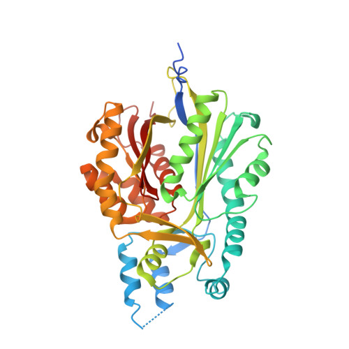
A novel tunnel in mycobacterial type III polyketide synthase reveals the structural basis for generating diverse metabolites

Sankaranarayanan, R.Saxena, P.Marathe, U.B.Gokhale, R.S.Shanmugam, V.M.Rukmini, R.

(2004) Nat Struct Mol Biol 11: 894-900

  • DOI: https://doi.org/10.1038/nsmb809
  • Primary Citation of Related Structures:  
    1TED, 1TEE

  • PubMed Abstract: 

    The superfamily of plant and bacterial type III polyketide synthases (PKSs) produces diverse metabolites with distinct biological functions. PKS18, a type III PKS from Mycobacterium tuberculosis, displays an unusual broad specificity for aliphatic long-chain acyl-coenzyme A (acyl-CoA) starter units (C(6)-C(20)) to produce tri- and tetraketide pyrones. The crystal structure of PKS18 reveals a 20 A substrate binding tunnel, hitherto unidentified in this superfamily of enzymes. This remarkable tunnel extends from the active site to the surface of the protein and is primarily generated by subtle changes of backbone dihedral angles in the core of the protein. Mutagenic studies combined with structure determination provide molecular insights into the structural elements that contribute to the chain length specificity of the enzyme. This first bacterial type III PKS structure underlines a fascinating example of the way in which subtle changes in protein architecture can generate metabolite diversity in nature.


  • Organizational Affiliation

    Centre for Cellular and Molecular Biology, Uppal Road, Hyderabad 500 007, India. sankar@ccmb.res.in


Macromolecules
Find similar proteins by:  (by identity cutoff)  |  3D Structure
Entity ID: 1
MoleculeChains Sequence LengthOrganismDetailsImage
pks18
A, B, C, D
393Mycobacterium tuberculosis H37RvMutation(s): 0 
Gene Names: pks18
EC: 2.3.1.74
UniProt
Find proteins for P9WPF1 (Mycobacterium tuberculosis (strain ATCC 25618 / H37Rv))
Explore P9WPF1 
Go to UniProtKB:  P9WPF1
Entity Groups  
Sequence Clusters30% Identity50% Identity70% Identity90% Identity95% Identity100% Identity
UniProt GroupP9WPF1
Sequence Annotations
Expand
  • Reference Sequence
Experimental Data & Validation

Experimental Data

  • Method: X-RAY DIFFRACTION
  • Resolution: 2.25 Å
  • R-Value Free: 0.265 
  • R-Value Work: 0.205 
  • R-Value Observed: 0.205 
  • Space Group: P 1
Unit Cell:
Length ( Å )Angle ( ˚ )
a = 59.914α = 108.23
b = 80.654β = 92.97
c = 99.612γ = 103.69
Software Package:
Software NamePurpose
CNSrefinement
DENZOdata reduction
SCALEPACKdata scaling
CCP4phasing

Structure Validation

View Full Validation Report


Ligand Structure Quality Assessment 


Entry History 

Deposition Data

Revision History  (Full details and data files)

  • Version 1.0: 2004-08-03
    Type: Initial release
  • Version 1.1: 2008-04-30
    Changes: Version format compliance
  • Version 1.2: 2011-07-13
    Changes: Source and taxonomy, Version format compliance
  • Version 1.3: 2023-10-25
    Changes: Data collection, Database references, Derived calculations, Refinement description