1T5E

The structure of MexA


Experimental Data Snapshot

  • Method: X-RAY DIFFRACTION
  • Resolution: 3.00 Å
  • R-Value Free: 0.285 
  • R-Value Work: 0.273 
  • R-Value Observed: 0.274 

wwPDB Validation   3D Report Full Report


This is version 2.0 of the entry. See complete history


Literature

Structure of the periplasmic component of a bacterial drug efflux pump

Higgins, M.K.Bokma, E.Koronakis, E.Hughes, C.Koronakis, V.

(2004) Proc Natl Acad Sci U S A 101: 9994-9999

  • DOI: https://doi.org/10.1073/pnas.0400375101
  • Primary Citation of Related Structures:  
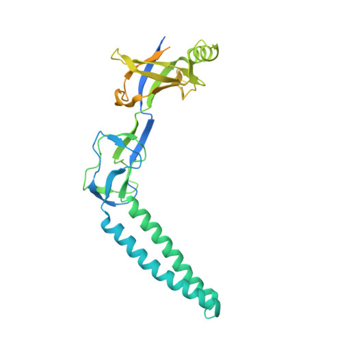
    1T5E

  • PubMed Abstract: 

    Multidrug resistance among Gram-negative bacteria is conferred by three-component membrane pumps that expel diverse antibiotics from the cell. These efflux pumps consist of an inner membrane transporter such as the AcrB proton antiporter, an outer membrane exit duct of the TolC family, and a periplasmic protein known as the adaptor. We present the x-ray structure of the MexA adaptor from the human pathogen Pseudomonas aeruginosa. The elongated molecule contains three linearly arranged subdomains; a 47-A-long alpha-helical hairpin, a lipoyl domain, and a six-stranded beta-barrel. In the crystal, hairpins of neighboring MexA monomers pack side-by-side to form twisted arcs. We discuss the implications of the packing of molecules within the crystal. On the basis of the structure and packing, we suggest a model for the key periplasmic interaction between the outer membrane channel and the adaptor protein in the assembled drug efflux pump.


  • Organizational Affiliation

    Department of Pathology, Cambridge University, Tennis Court Road, Cambridge CB2 1QP, United Kingdom. mkh20@hermes.cam.ac.uk


Macromolecules
Find similar proteins by:  (by identity cutoff)  |  3D Structure
Entity ID: 1
MoleculeChains Sequence LengthOrganismDetailsImage
Multidrug resistance protein mexA
A, B, C, D, E
A, B, C, D, E, F, G, H, I, J, K, L, M
360Pseudomonas aeruginosaMutation(s): 0 
Gene Names: MEXAPA0425
Membrane Entity: Yes 
UniProt
Find proteins for P52477 (Pseudomonas aeruginosa (strain ATCC 15692 / DSM 22644 / CIP 104116 / JCM 14847 / LMG 12228 / 1C / PRS 101 / PAO1))
Explore P52477 
Go to UniProtKB:  P52477
Entity Groups  
Sequence Clusters30% Identity50% Identity70% Identity90% Identity95% Identity100% Identity
UniProt GroupP52477
Sequence Annotations
Expand
  • Reference Sequence
Small Molecules
Ligands 2 Unique
IDChains Name / Formula / InChI Key2D Diagram3D Interactions
GOL
Query on GOL

Download Ideal Coordinates CCD File 
N [auth A],
O [auth B],
W [auth J],
Z [auth M]
GLYCEROL
C3 H8 O3
PEDCQBHIVMGVHV-UHFFFAOYSA-N
3GR
Query on 3GR

Download Ideal Coordinates CCD File 
P [auth C]
Q [auth D]
R [auth E]
S [auth F]
T [auth G]
P [auth C],
Q [auth D],
R [auth E],
S [auth F],
T [auth G],
U [auth H],
V [auth I],
X [auth K],
Y [auth L]
D-Glyceraldehyde
C3 H6 O3
MNQZXJOMYWMBOU-VKHMYHEASA-N
Experimental Data & Validation

Experimental Data

  • Method: X-RAY DIFFRACTION
  • Resolution: 3.00 Å
  • R-Value Free: 0.285 
  • R-Value Work: 0.273 
  • R-Value Observed: 0.274 
  • Space Group: P 1 21 1
Unit Cell:
Length ( Å )Angle ( ˚ )
a = 130.551α = 90
b = 183.585β = 107.38
c = 213.314γ = 90
Software Package:
Software NamePurpose
MOSFLMdata reduction
SCALAdata scaling
SHARPphasing
CNSrefinement
CCP4data scaling

Structure Validation

View Full Validation Report


Entry History 

Deposition Data

Revision History  (Full details and data files)

  • Version 1.0: 2004-05-18
    Type: Initial release
  • Version 1.1: 2008-04-30
    Changes: Version format compliance
  • Version 1.2: 2011-07-13
    Changes: Non-polymer description, Version format compliance
  • Version 2.0: 2020-07-08
    Changes: Atomic model, Data collection, Derived calculations, Structure summary