1SUM

Crystal structure of a hypothetical protein at 2.0 A resolution


Experimental Data Snapshot

  • Method: X-RAY DIFFRACTION
  • Resolution: 2.00 Å
  • R-Value Free: 0.255 
  • R-Value Work: 0.217 
  • R-Value Observed: 0.220 

wwPDB Validation   3D Report Full Report


This is version 1.3 of the entry. See complete history


Literature

Crystal structure of a PhoU protein homologue: a new class of metalloprotein containing multinuclear iron clusters.

Liu, J.Lou, Y.Yokota, H.Adams, P.D.Kim, R.Kim, S.H.

(2005) J Biol Chem 280: 15960-15966

  • DOI: https://doi.org/10.1074/jbc.M414117200
  • Primary Citation of Related Structures:  
    1SUM

  • PubMed Abstract: 

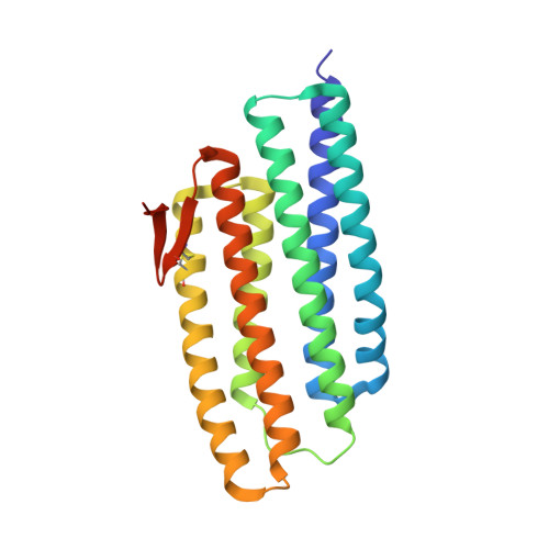
    PhoU proteins are known to play a role in the regulation of phosphate uptake. In Thermotoga maritima, two PhoU homologues have been identified bioinformatically. Here we report the crystal structure of one of the PhoU homologues at 2.0 A resolution. The structure of the PhoU protein homologue contains a highly symmetric new structural fold composed of two repeats of a three-helix bundle. The structure unexpectedly revealed a trinuclear and a tetranuclear iron cluster that were found to be bound on the surface. Each of the two multinuclear iron clusters is coordinated by a conserved E(D)XXXD motif pair. Our structure reveals a new class of metalloprotein containing multinuclear iron clusters. The possible functional implication based on the structure are discussed.


  • Organizational Affiliation

    Berkeley Structural Genomics Center, Lawrence Berkeley National Laboratory, Berkeley, California 94720, USA.


Macromolecules
Find similar proteins by:  (by identity cutoff)  |  3D Structure
Entity ID: 1
MoleculeChains Sequence LengthOrganismDetailsImage
Phosphate transport system protein phoU homolog 2A [auth B]235Thermotoga maritimaMutation(s): 0 
UniProt
Find proteins for Q9X256 (Thermotoga maritima (strain ATCC 43589 / DSM 3109 / JCM 10099 / NBRC 100826 / MSB8))
Explore Q9X256 
Go to UniProtKB:  Q9X256
Entity Groups  
Sequence Clusters30% Identity50% Identity70% Identity90% Identity95% Identity100% Identity
UniProt GroupQ9X256
Sequence Annotations
Expand
  • Reference Sequence
Small Molecules
Ligands 4 Unique
IDChains Name / Formula / InChI Key2D Diagram3D Interactions
PO3
Query on PO3

Download Ideal Coordinates CCD File 
K [auth B],
L [auth B]
PHOSPHITE ION
O3 P
AQSJGOWTSHOLKH-UHFFFAOYSA-N
NI
Query on NI

Download Ideal Coordinates CCD File 
I [auth B]NICKEL (II) ION
Ni
VEQPNABPJHWNSG-UHFFFAOYSA-N
FE
Query on FE

Download Ideal Coordinates CCD File 
B
C [auth B]
D [auth B]
E [auth B]
F [auth B]
B,
C [auth B],
D [auth B],
E [auth B],
F [auth B],
G [auth B],
H [auth B]
FE (III) ION
Fe
VTLYFUHAOXGGBS-UHFFFAOYSA-N
CA
Query on CA

Download Ideal Coordinates CCD File 
J [auth B]CALCIUM ION
Ca
BHPQYMZQTOCNFJ-UHFFFAOYSA-N
Experimental Data & Validation

Experimental Data

  • Method: X-RAY DIFFRACTION
  • Resolution: 2.00 Å
  • R-Value Free: 0.255 
  • R-Value Work: 0.217 
  • R-Value Observed: 0.220 
  • Space Group: P 31 2 1
Unit Cell:
Length ( Å )Angle ( ˚ )
a = 90.677α = 90
b = 90.677β = 90
c = 45.255γ = 120
Software Package:
Software NamePurpose
HKL-2000data collection
SCALEPACKdata scaling
PHENIXmodel building
REFMACrefinement
HKL-2000data reduction
PHENIXphasing

Structure Validation

View Full Validation Report


Entry History 

Deposition Data

Revision History  (Full details and data files)

  • Version 1.0: 2004-08-24
    Type: Initial release
  • Version 1.1: 2008-04-29
    Changes: Version format compliance
  • Version 1.2: 2011-07-13
    Changes: Advisory, Derived calculations, Version format compliance
  • Version 1.3: 2017-10-11
    Changes: Refinement description