Spectroscopic studies of a phosphoinositide-binding peptide from gelsolin: behavior in solutions of mixed solvent and anionic micelles.
Xian, W., Vegners, R., Janmey, P.A., Braunlin, W.H.(1995) Biophys J 69: 2695-2702
- PubMed: 8599675
- DOI: https://doi.org/10.1016/S0006-3495(95)80140-2
- Primary Citation of Related Structures:
1SOL - PubMed Abstract:
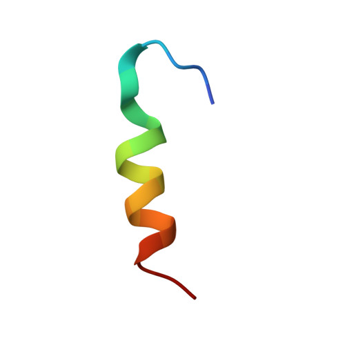
The peptide G(150-169) corresponds to a phosphatidylinositol 4,5-bisphosphate (PIP2) and filamentous actin (F-actin) binding site on gelsolin (residues 150-169, with the sequence KHVVPNEVVVQRLFQVKGRR). The conformation of this peptide in trifluoroethanol (TFE) aqueous solution was determined by 1H nuclear magnetic resonance as the first step toward understanding the structural aspects of the interaction of G(150-169) and PIP2. The circular dichroism experiments show that G(150-169) adopts a predominantly alpha-helical form in both 50% TFE aqueous solution and in the presence of PIP2 micelles, therefore establishing a connection between the two conformations. 1H nuclear magnetic resonance experiments of G(150-169) in TFE co-solvent show that the helical region extends from Pro-154 to Lys-166. The amphiphilic nature of this helical structure may be the key to understanding the binding of the peptide to lipids. Sodium dodecyl sulfate micelle solution is used as a model for anionic lipid environments. Preliminary studies of the conformation of G(150-169) in sodium dodecyl sulfate micelle solution show that the peptide forms an alpha-helix similar to but with some structural differences from that in TFE co-solvent. Fluorescence experiments provide evidence of peptide clustering over a narrow range of peptide/PIP2 ratios, which is potentially relevant to the biological function of PIP2.
Organizational Affiliation:
Department of Chemistry, University of Nebraska-Lincoln 68588-0304, USA.