1SKO

MP1-p14 Complex


Experimental Data Snapshot

  • Method: X-RAY DIFFRACTION
  • Resolution: 2.00 Å
  • R-Value Free: 0.271 
  • R-Value Work: 0.201 
  • R-Value Observed: 0.205 

wwPDB Validation   3D Report Full Report


This is version 1.3 of the entry. See complete history


Literature

The structure of the MAP kinase scaffold MP1 bound to its partner p14: a complex with a critical role in endosomal MAP kinase signaling

Lunin, V.V.Munger, C.Wagner, J.Ye, Z.Cygler, M.Sacher, M.

(2004) J Biol Chem 279: 23422-23430

  • DOI: https://doi.org/10.1074/jbc.M401648200
  • Primary Citation of Related Structures:  
    1SKO

  • PubMed Abstract: 

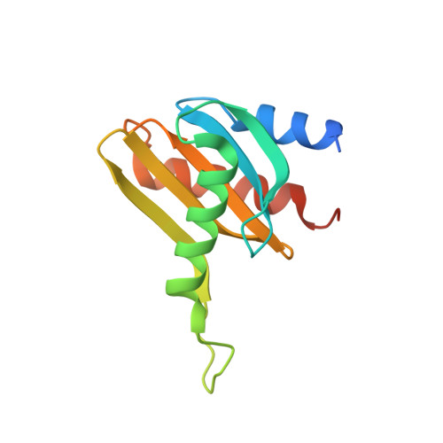
    Scaffold proteins of the mitogen-activated protein kinase (MAPK) pathway have been proposed to form an active signaling module and enhance the specificity of the transduced signal. Here, we report a 2-A resolution structure of the MAPK scaffold protein MP1 in a complex with its partner protein, p14, that localizes the complex to late endosomes. The structures of these two proteins are remarkably similar, with a five-stranded beta-sheet flanked on either side by a total of three helices. The proteins form a heterodimer in solution and interact mainly through the edge beta-strand in each protein to generate a 10-stranded beta-sheet core. Both proteins also share structural similarity with the amino-terminal regulatory domains of the membrane trafficking proteins, sec22b and Ykt6p, as well as with sedlin (a component of a Golgi-associated membrane-trafficking complex) and the sigma2 and amino-terminal portion of the mu2 subunits of the clathrin adaptor complex AP2. Because neither MP1 nor p14 have been implicated in membrane traffic, we propose that the similar protein folds allow these relatively small proteins to be involved in multiple and simultaneous protein-protein interactions. Mapping of highly conserved, surface-exposed residues on MP1 and p14 provided insight into the potential sites of binding of the signaling kinases MEK1 and ERK1 to this complex, as well as the areas potentially involved in other protein-protein interactions.


  • Organizational Affiliation

    Biotechnology Research Institute, National Research Council of Canada, Montréal, Québec H4P 2R2, Canada.


Macromolecules
Find similar proteins by:  (by identity cutoff)  |  3D Structure
Entity ID: 1
MoleculeChains Sequence LengthOrganismDetailsImage
Mitogen-activated protein kinase kinase 1 interacting protein 1143Homo sapiensMutation(s): 0 
Gene Names: MAP2K1IP1
UniProt & NIH Common Fund Data Resources
Find proteins for Q9UHA4 (Homo sapiens)
Explore Q9UHA4 
Go to UniProtKB:  Q9UHA4
PHAROS:  Q9UHA4
GTEx:  ENSG00000109270 
Entity Groups  
Sequence Clusters30% Identity50% Identity70% Identity90% Identity95% Identity100% Identity
UniProt GroupQ9UHA4
Sequence Annotations
Expand
  • Reference Sequence
Find similar proteins by:  (by identity cutoff)  |  3D Structure
Entity ID: 2
MoleculeChains Sequence LengthOrganismDetailsImage
Late endosomal/lysosomal Mp1 interacting protein130Mus musculusMutation(s): 0 
Gene Names: MAPBPIP
UniProt
Find proteins for Q9JHS3 (Mus musculus)
Explore Q9JHS3 
Go to UniProtKB:  Q9JHS3
Entity Groups  
Sequence Clusters30% Identity50% Identity70% Identity90% Identity95% Identity100% Identity
UniProt GroupQ9JHS3
Sequence Annotations
Expand
  • Reference Sequence
Experimental Data & Validation

Experimental Data

  • Method: X-RAY DIFFRACTION
  • Resolution: 2.00 Å
  • R-Value Free: 0.271 
  • R-Value Work: 0.201 
  • R-Value Observed: 0.205 
  • Space Group: P 21 21 21
Unit Cell:
Length ( Å )Angle ( ˚ )
a = 50.974α = 90
b = 64.193β = 90
c = 73.28γ = 90
Software Package:
Software NamePurpose
REFMACrefinement
HKL-2000data reduction
SCALEPACKdata scaling
SOLVEphasing

Structure Validation

View Full Validation Report


Entry History 

Deposition Data

Revision History  (Full details and data files)

  • Version 1.0: 2004-06-01
    Type: Initial release
  • Version 1.1: 2008-04-29
    Changes: Version format compliance
  • Version 1.2: 2011-07-13
    Changes: Version format compliance
  • Version 1.3: 2024-02-14
    Changes: Data collection, Database references