1S5V

Crystal Structure Analysis of a mutant of DIHYDRODIPICOLINATE SYNTHASE--residue Tyr107 to Phe107


Experimental Data Snapshot

  • Method: X-RAY DIFFRACTION
  • Resolution: 2.35 Å
  • R-Value Free: 0.232 
  • R-Value Work: 0.195 
  • R-Value Observed: 0.197 

wwPDB Validation   3D Report Full Report


This is version 1.4 of the entry. See complete history


Literature

The Crystal Structure of Three Site-directed Mutants of Escherichia coli Dihydrodipicolinate Synthase: Further Evidence for a Catalytic Triad

Dobson, R.C.J.Valegard, K.Gerrard, J.A.

(2004) J Mol Biol 338: 329-339

  • DOI: https://doi.org/10.1016/j.jmb.2004.02.060
  • Primary Citation of Related Structures:  
    1S5T, 1S5V, 1S5W

  • PubMed Abstract: 

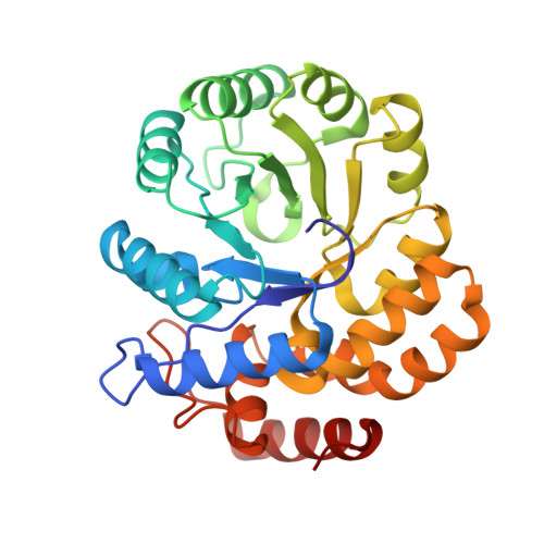
    Dihydrodipicolinate synthase (DHDPS, EC 4.2.1.52) catalyses the branchpoint reaction of lysine biosynthesis in plants and microbes: the condensation of (S)-aspartate-beta-semialdehyde and pyruvate. The crystal structure of wild-type DHDPS has been published to 2.5A, revealing a tetrameric molecule comprised of four identical (beta/alpha)(8)-barrels, each containing one active site. Previous workers have hypothesised that the catalytic mechanism of the enzyme involves a catalytic triad of amino acid residues, Tyr133, Thr44 and Tyr107, which provide a proton shuttle to transport protons from the active site to solvent. We have tested this hypothesis using site-directed mutagenesis to produce three mutant enzymes: DHDPS-Y133F, DHDPS-T44V and DHDPS-Y107F. Each of these mutants has substantially reduced activity, consistent with the catalytic triad hypothesis. We have determined each mutant crystal structure to at least 2.35A resolution and compared the structures to the wild-type enzyme. All mutant enzymes crystallised in the same space group as the wild-type form and only minor differences in structure are observed. These results suggest that the catalytic triad is indeed in operation in wild-type DHDPS.


  • Organizational Affiliation

    Molecular Biophysics, Department of Cell and Molecular Biology, Uppsala University, Box 596, S-751 24 Uppsala, Sweden.


Macromolecules
Find similar proteins by:  (by identity cutoff)  |  3D Structure
Entity ID: 1
MoleculeChains Sequence LengthOrganismDetailsImage
Dihydrodipicolinate synthase
A, B
292Escherichia coliMutation(s): 1 
Gene Names: DAPA
EC: 4.2.1.52
UniProt
Find proteins for P0A6L2 (Escherichia coli (strain K12))
Explore P0A6L2 
Go to UniProtKB:  P0A6L2
Entity Groups  
Sequence Clusters30% Identity50% Identity70% Identity90% Identity95% Identity100% Identity
UniProt GroupP0A6L2
Sequence Annotations
Expand
  • Reference Sequence
Experimental Data & Validation

Experimental Data

  • Method: X-RAY DIFFRACTION
  • Resolution: 2.35 Å
  • R-Value Free: 0.232 
  • R-Value Work: 0.195 
  • R-Value Observed: 0.197 
  • Space Group: P 31 2 1
Unit Cell:
Length ( Å )Angle ( ˚ )
a = 120.888α = 90
b = 120.888β = 90
c = 110.202γ = 120
Software Package:
Software NamePurpose
REFMACrefinement
DENZOdata reduction
SCALEPACKdata scaling

Structure Validation

View Full Validation Report


Entry History 

Deposition Data

Revision History  (Full details and data files)

  • Version 1.0: 2004-04-27
    Type: Initial release
  • Version 1.1: 2008-04-29
    Changes: Version format compliance
  • Version 1.2: 2011-07-13
    Changes: Derived calculations, Version format compliance
  • Version 1.3: 2021-11-10
    Changes: Database references
  • Version 1.4: 2023-10-25
    Changes: Data collection, Refinement description