Evolution of Enzymatic Activity in the Enolase Superfamily: Structural and Mutagenic Studies of the Mechanism of the Reaction Catalyzed by o-Succinylbenzoate Synthase from Escherichia coli
Klenchin, V.A., Taylor Ringia, E.A., Gerlt, J.A., Rayment, I.(2003) Biochemistry 42: 14427-14433
- PubMed: 14661953
- DOI: https://doi.org/10.1021/bi035545v
- Primary Citation of Related Structures:
1R6W - PubMed Abstract:
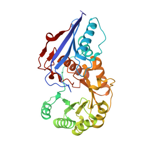
o-Succinylbenzoate synthase (OSBS) from Escherichia coli, a member of the enolase superfamily, catalyzes an exergonic dehydration reaction in the menaquinone biosynthetic pathway in which 2-succinyl-6-hydroxy-2,4-cyclohexadiene-1-carboxylate (SHCHC) is converted to 4-(2'-carboxyphenyl)-4-oxobutyrate (o-succinylbenzoate or OSB). Our previous structural studies of the Mg(2+).OSB complex established that OSBS is a member of the muconate lactonizing enzyme subgroup of the superfamily: the essential Mg(2+) is coordinated to carboxylate ligands at the ends of the third, fourth, and fifth beta-strands of the (beta/alpha)(7)beta-barrel catalytic domain, and the OSB product is located between the Lys 133 at the end of the second beta-strand and the Lys 235 at the end of the sixth beta-strand [Thompson, T. B., Garrett, J. B., Taylor, E. A, Meganathan, R., Gerlt, J. A., and Rayment, I. (2000) Biochemistry 39, 10662-76]. Both Lys 133 and Lys 235 were separately replaced with Ala, Ser, and Arg residues; all six mutants displayed no detectable catalytic activity. The structure of the Mg(2+).SHCHC complex of the K133R mutant has been solved at 1.62 A resolution by molecular replacement starting from the structure of the Mg(2+).OSB complex. This establishes the absolute configuration of SHCHC: the C1-carboxylate and the C6-OH leaving group are in a trans orientation, requiring that the dehydration proceed via a syn stereochemical course. The side chain of Arg 133 is pointed out of the active site so that it cannot function as a general base, whereas in the wild-type enzyme complexed with Mg(2+).OSB, the side chain of Lys 133 is appropriately positioned to function as the only acid/base catalyst in the syn dehydration. The epsilon-ammonium group of Lys 235 forms a cation-pi interaction with the cyclohexadienyl moiety of SHCHC, suggesting that Lys 235 also stabilizes the enediolate anion intermediate in the syn dehydration via a similar interaction.
Organizational Affiliation:
Department of Biochemistry, University of Wisconsin, Madison, Wisconsin 53705, USA.