1R6N

HPV11 E2 TAD complex crystal structure


Experimental Data Snapshot

  • Method: X-RAY DIFFRACTION
  • Resolution: 2.40 Å
  • R-Value Free: 0.266 
  • R-Value Work: 0.209 
  • R-Value Observed: 0.209 

wwPDB Validation   3D Report Full Report


Ligand Structure Quality Assessment 


This is version 1.5 of the entry. See complete history


Literature

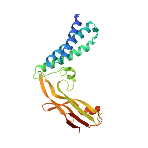
Crystal Structure of the E2 Transactivation Domain of Human Papillomavirus Type 11 Bound to a Protein Interaction Inhibitor

Wang, Y.Coulombe, R.Cameron, D.R.Thauvette, L.Massariol, M.-J.Amon, L.M.Fink, D.Titolo, S.Welchner, E.Yoakim, C.Archambault, J.White, P.W.

(2004) J Biol Chem 279: 6976-6985

  • DOI: https://doi.org/10.1074/jbc.M311376200
  • Primary Citation of Related Structures:  
    1R6K, 1R6N

  • PubMed Abstract: 

    Interaction between the E2 protein and E1 helicase of human papillomaviruses (HPVs) is essential for the initiation of viral DNA replication. We recently described a series of small molecules that bind to the N-terminal transactivation domain (TAD) of HPV type 11 E2 and inhibits its interaction with E1 in vitro and in cellular assays. Here we report the crystal structures of both the HPV11 TAD and of a complex between this domain and an inhibitor, at 2.5- and 2.4-A resolution, respectively. The HPV11 TAD structure is very similar to that of the analogous domain of HPV16. Inhibitor binding caused no significant alteration of the protein backbone, but movements of several amino acid side chains at the binding site, in particular those of Tyr-19, His-32, Leu-94, and Glu-100, resulted in the formation of a deep hydrophobic pocket that accommodates the indandione moiety of the inhibitor. Mutational analysis provides functional evidence for specific interactions between Tyr-19 and E1 and between His-32 and the inhibitor. A second inhibitor molecule is also present at the binding pocket. Although evidence is presented that this second molecule makes only weak interactions with the protein and is likely an artifact of crystallization, its presence defines additional regions of the binding pocket that could be exploited to design more potent inhibitors.


  • Organizational Affiliation

    Department of Medicinal Chemistry, Boehringer Ingelheim Pharmaceuticals Inc, Ridgefield, CT 06877, USA.


Macromolecules
Find similar proteins by:  (by identity cutoff)  |  3D Structure
Entity ID: 1
MoleculeChains Sequence LengthOrganismDetailsImage
HPV11 REGULATORY PROTEIN E2211human papillomavirus 11Mutation(s): 0 
Gene Names: E2
UniProt
Find proteins for P04015 (Human papillomavirus 11)
Explore P04015 
Go to UniProtKB:  P04015
Entity Groups  
Sequence Clusters30% Identity50% Identity70% Identity90% Identity95% Identity100% Identity
UniProt GroupP04015
Sequence Annotations
Expand
  • Reference Sequence
Small Molecules
Ligands 3 Unique
IDChains Name / Formula / InChI Key2D Diagram3D Interactions
434
Query on 434

Download Ideal Coordinates CCD File 
B [auth A],
C [auth A]
SPIRO[3-CARBOXY-4-{(4-[1,2,3]THIADIAZOL-4-YL-PHENYL)-AMINO-CARBONYL} -5-[3,4-DICHLORO-PHENYL]-TETRAHYDROFURAN-2,2'-5-METHYL-INDAN-1,3-DIONE]
C29 H19 Cl2 N3 O6 S
KNRVCCXHLSHTFW-HGPRPZRGSA-N
ALQ
Query on ALQ

Download Ideal Coordinates CCD File 
D [auth A]2-METHYL-PROPIONIC ACID
C4 H8 O2
KQNPFQTWMSNSAP-UHFFFAOYSA-N
DMS
Query on DMS

Download Ideal Coordinates CCD File 
E [auth A]DIMETHYL SULFOXIDE
C2 H6 O S
IAZDPXIOMUYVGZ-UHFFFAOYSA-N
Binding Affinity Annotations 
IDSourceBinding Affinity
434 Binding MOAD:  1R6N Kd: 40 (nM) from 1 assay(s)
PDBBind:  1R6N Kd: 40 (nM) from 1 assay(s)
Experimental Data & Validation

Experimental Data

  • Method: X-RAY DIFFRACTION
  • Resolution: 2.40 Å
  • R-Value Free: 0.266 
  • R-Value Work: 0.209 
  • R-Value Observed: 0.209 
  • Space Group: P 41
Unit Cell:
Length ( Å )Angle ( ˚ )
a = 60.456α = 90
b = 60.456β = 90
c = 82.526γ = 90
Software Package:
Software NamePurpose
DENZOdata reduction
SCALEPACKdata scaling
CNSrefinement
CNSphasing

Structure Validation

View Full Validation Report


Ligand Structure Quality Assessment 


Entry History 

Deposition Data

Revision History  (Full details and data files)

  • Version 1.0: 2004-02-24
    Type: Initial release
  • Version 1.1: 2008-04-29
    Changes: Version format compliance
  • Version 1.2: 2011-07-13
    Changes: Version format compliance
  • Version 1.3: 2018-01-24
    Changes: Database references
  • Version 1.4: 2022-12-21
    Changes: Database references, Derived calculations
  • Version 1.5: 2023-09-20
    Changes: Data collection, Refinement description