1PYG

STRUCTURAL BASIS FOR THE ACTIVATION OF GLYCOGEN PHOSPHORYLASE B BY ADENOSINE MONOPHOSPHATE


Experimental Data Snapshot

  • Method: X-RAY DIFFRACTION
  • Resolution: 2.87 Å
  • R-Value Work: 0.185 

wwPDB Validation   3D Report Full Report


This is version 1.2 of the entry. See complete history


Literature

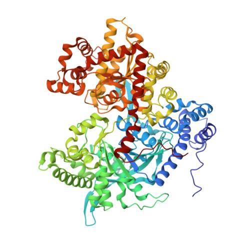
Structural basis for the activation of glycogen phosphorylase b by adenosine monophosphate.

Sprang, S.R.Withers, S.G.Goldsmith, E.J.Fletterick, R.J.Madsen, N.B.

(1991) Science 254: 1367-1371

  • DOI: https://doi.org/10.1126/science.1962195
  • Primary Citation of Related Structures:  
    1PYG

  • PubMed Abstract: 

    The three-dimensional structure of the activated state of glycogen phosphorylase (GP) as induced by adenosine monophosphate (AMP) has been determined from crystals of pyridoxalpyrophosphoryl-GP. The same quaternary changes relative to the inactive conformation as those induced by phosphorylation are induced by AMP, although the two regulatory signals function through different local structural mechanisms. Moreover, previous descriptions of the phosphorylase active state have been extended by demonstrating that, on activation, the amino- and carboxyl-terminal domains of GP rotate apart by 5 degrees, thereby increasing access of substrates to the catalytic site. The structure also reveals previously unobserved interactions with the nucleotide that accounts for the specificity of the nucleotide binding site for AMP in preference to inosine monophosphate.


  • Organizational Affiliation

    Howard Hughes Medical Institute, University of Texas Southwestern Medical Center, Dallas 75235-9050.


Macromolecules
Find similar proteins by:  (by identity cutoff)  |  3D Structure
Entity ID: 1
MoleculeChains Sequence LengthOrganismDetailsImage
GLYCOGEN PHOSPHORYLASE B
A, B, C, D
842Oryctolagus cuniculusMutation(s): 0 
EC: 2.4.1.1
UniProt
Find proteins for P00489 (Oryctolagus cuniculus)
Explore P00489 
Go to UniProtKB:  P00489
Entity Groups  
Sequence Clusters30% Identity50% Identity70% Identity90% Identity95% Identity100% Identity
UniProt GroupP00489
Sequence Annotations
Expand
  • Reference Sequence
Experimental Data & Validation

Experimental Data

  • Method: X-RAY DIFFRACTION
  • Resolution: 2.87 Å
  • R-Value Work: 0.185 
  • Space Group: P 21 21 2
Unit Cell:
Length ( Å )Angle ( ˚ )
a = 169.9α = 90
b = 209.9β = 90
c = 123.4γ = 90
Software Package:
Software NamePurpose
X-PLORmodel building
X-PLORrefinement
X-PLORphasing

Structure Validation

View Full Validation Report


Entry History 

Deposition Data

  • Released Date: 1994-01-31 
  • Deposition Author(s): Sprang, S.

Revision History  (Full details and data files)

  • Version 1.0: 1994-01-31
    Type: Initial release
  • Version 1.1: 2008-03-03
    Changes: Version format compliance
  • Version 1.2: 2011-07-13
    Changes: Derived calculations, Version format compliance