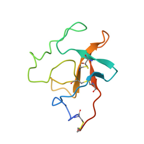
Kringle-kringle interactions in multimer kringle structures.
Padmanabhan, K., Wu, T.P., Ravichandran, K.G., Tulinsky, A.(1994) Protein Sci 3: 898-910
- PubMed: 8069221
- DOI: https://doi.org/10.1002/pro.5560030605
- Primary Citation of Related Structures:
1PMK, 1PML - PubMed Abstract:
The crystal structure of a monoclinic form of human plasminogen kringle 4 (PGK4) has been solved by molecular replacement using the orthorthombic structure as a model and it has been refined by restrained least-squares methods to an R factor of 16.4% at 2.25 A resolution. The X-PLOR structure of kringle 2 of tissue plasminogen activator (t-PAK2) has been refined further using PROFFT (R = 14.5% at 2.38 A resolution). The PGK4 structure has 2 and t-PAK2 has 3 independent molecules in the asymmetric unit. There are 5 different noncrystallographic symmetry "dimers" in PGK4. Three make extensive kringle-kringle interactions related by noncrystallographic 2(1) screw axes without blocking the lysine binding site. Such associations may occur in multikringle structures such as prothrombin, hepatocyte growth factor, plasminogen (PG), and apolipoprotein [a]. The t-PAK2 structure also has noncrystallographic screw symmetry (3(1)) and mimics fibrin binding mode by having lysine of one molecule interacting electrostatically with the lysine binding site of another kringle. This ligand-like binding interaction may be important in kringle-kringle interactions involving non-lysine binding kringles with lysine or pseudo-lysine binding sites. Electrostatic intermolecular interactions involving the lysine binding site are also found in the crystal structures of PGK1 and orthorhombic PGK4. Anions associate with the cationic centers of these and t-PAK2 that appear to be more than occasional components of lysine binding site regions.
Organizational Affiliation:
Department of Chemistry, Michigan State University, East Lansing 48824.