1PIE

Crystal Structure of Lactococcus lactis Galactokinase Complexed with Galactose


Experimental Data Snapshot

  • Method: X-RAY DIFFRACTION
  • Resolution: 2.10 Å
  • R-Value Free: 0.266 
  • R-Value Work: 0.199 
  • R-Value Observed: 0.204 

wwPDB Validation   3D Report Full Report


Ligand Structure Quality Assessment 


This is version 1.4 of the entry. See complete history


Literature

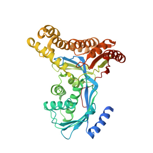
Molecular Structure of Galactokinase

Thoden, J.B.Holden, H.M.

(2003) J Biol Chem 278: 33305-33311

  • DOI: https://doi.org/10.1074/jbc.M304789200
  • Primary Citation of Related Structures:  
    1PIE

  • PubMed Abstract: 

    Galactokinase plays a key role in normal galactose metabolism by catalyzing the ATP-dependent phosphorylation of alpha-D-galactose to galactose 1-phosphate. In humans, mutations in the galactokinase gene can lead to the diseased state referred to as Type II galactosemia. Here we describe the three-dimensional structure of galactokinase from Lactococcus lactis determined to 2.1-A resolution. As expected from amino acid sequence alignments, galactokinase adopts a similar topology to that observed for members of the GHMP superfamily. The N-terminal domain is characterized by a five-stranded mixed beta-sheet while the C-terminal motif is dominated by two distinct four-stranded anti-parallel beta-sheets. The structure was solved in the presence of alpha-D-galactose and inorganic phosphate. These ligands are wedged between the N- and C-terminal domains. Amino acid side chains responsible for anchoring the sugar ligand to the protein include Arg36, Glu42, Asp45, Asp183, and Tyr233. Both Arg36 and Asp183 are strictly conserved in the amino acid sequences available in the literature thus far for galactokinases. Interestingly, the carboxylate side chain of Asp183 is positioned within 3.5 A of the C-1 hydroxyl group of galactose, whereas the guanidinium group of Arg36 is situated between both the C-1 hydroxyl group and the inorganic phosphate. Most likely these residues play key roles in catalysis. The structure of galactokinase described here serves as a model for understanding the functional consequences of point mutations known to result in Type II galactosemia in humans.


  • Organizational Affiliation

    Department of Biochemistry, University of Wisconsin, Madison, Wisconsin 53706, USA.


Macromolecules
Find similar proteins by:  (by identity cutoff)  |  3D Structure
Entity ID: 1
MoleculeChains Sequence LengthOrganismDetailsImage
Galactokinase419Lactococcus lactisMutation(s): 0 
Gene Names: GALK
EC: 2.7.1.6
UniProt
Find proteins for Q9R7D7 (Lactococcus lactis subsp. lactis (strain IL1403))
Explore Q9R7D7 
Go to UniProtKB:  Q9R7D7
Entity Groups  
Sequence Clusters30% Identity50% Identity70% Identity90% Identity95% Identity100% Identity
UniProt GroupQ9R7D7
Sequence Annotations
Expand
  • Reference Sequence
Small Molecules
Ligands 2 Unique
IDChains Name / Formula / InChI Key2D Diagram3D Interactions
GLA
Query on GLA

Download Ideal Coordinates CCD File 
B [auth A]alpha-D-galactopyranose
C6 H12 O6
WQZGKKKJIJFFOK-PHYPRBDBSA-N
PO4
Query on PO4

Download Ideal Coordinates CCD File 
C [auth A]PHOSPHATE ION
O4 P
NBIIXXVUZAFLBC-UHFFFAOYSA-K
Experimental Data & Validation

Experimental Data

  • Method: X-RAY DIFFRACTION
  • Resolution: 2.10 Å
  • R-Value Free: 0.266 
  • R-Value Work: 0.199 
  • R-Value Observed: 0.204 
  • Space Group: I 2 3
Unit Cell:
Length ( Å )Angle ( ˚ )
a = 143.4α = 90
b = 143.4β = 90
c = 143.4γ = 90
Software Package:
Software NamePurpose
d*TREKdata scaling
HKL-2000data reduction
SOLVEphasing
TNTrefinement
d*TREKdata reduction
HKL-2000data scaling

Structure Validation

View Full Validation Report


Ligand Structure Quality Assessment 


Entry History 

Deposition Data

Revision History  (Full details and data files)

  • Version 1.0: 2003-09-23
    Type: Initial release
  • Version 1.1: 2008-04-29
    Changes: Version format compliance
  • Version 1.2: 2011-07-13
    Changes: Version format compliance
  • Version 1.3: 2020-07-29
    Type: Remediation
    Reason: Carbohydrate remediation
    Changes: Data collection, Database references, Derived calculations, Structure summary
  • Version 1.4: 2024-02-14
    Changes: Data collection, Database references, Structure summary