1P9H

CRYSTAL STRUCTURE OF THE COLLAGEN-BINDING DOMAIN OF YERSINIA ADHESIN YadA


Experimental Data Snapshot

  • Method: X-RAY DIFFRACTION
  • Resolution: 1.55 Å
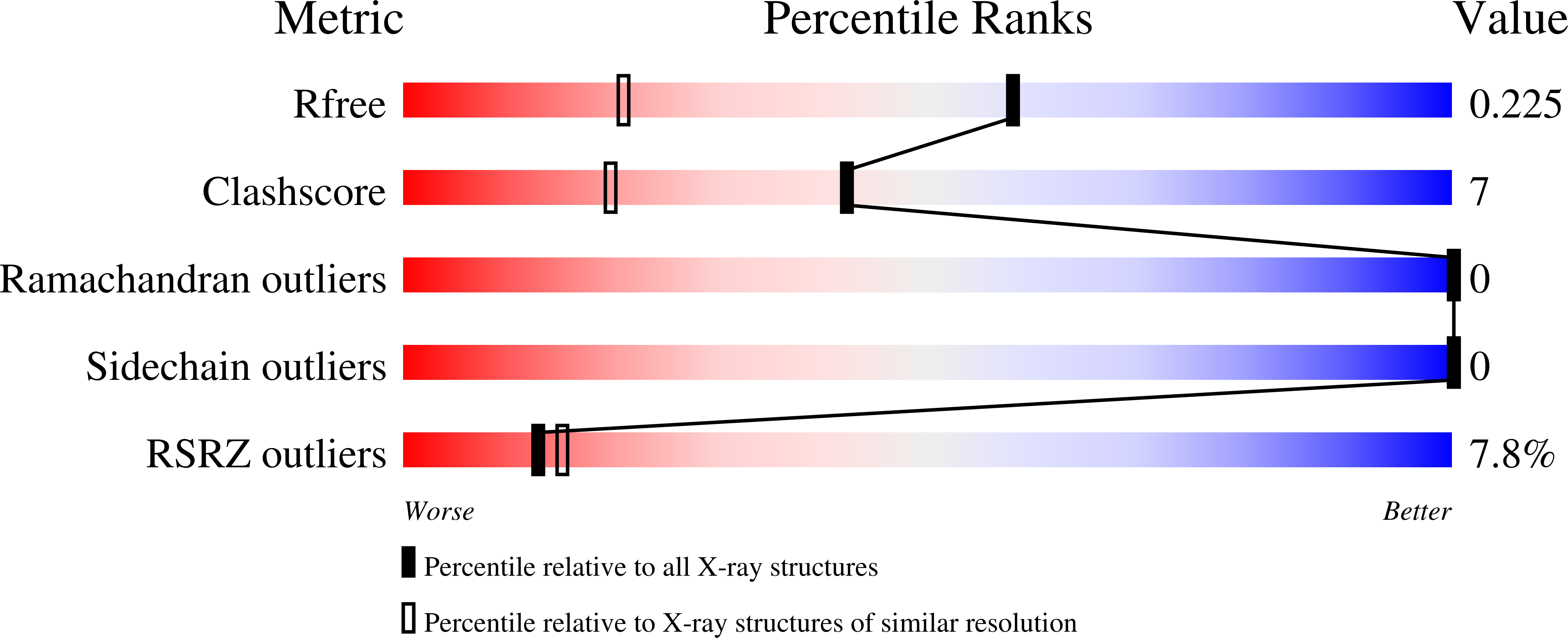
  • R-Value Free: 0.204 
  • R-Value Work: 0.196 

wwPDB Validation   3D Report Full Report


This is version 1.3 of the entry. See complete history


Literature

The Yersinia adhesin YadA collagen-binding domain structure is a novel left-handed parallel beta-roll.

Nummelin, H.Merckel, M.C.Leo, J.C.Lankinen, H.Skurnik, M.Goldman, A.

(2004) EMBO J 23: 701-711

  • DOI: https://doi.org/10.1038/sj.emboj.7600100
  • Primary Citation of Related Structures:  
    1P9H

  • PubMed Abstract: 

    The crystal structure of the recombinant collagen-binding domain of Yersinia adhesin YadA from Yersinia enterocolitica serotype O:3 was solved at 1.55 A resolution. The trimeric structure is composed of head and neck regions, and the collagen binding head region is a novel nine-coiled left-handed parallel beta-roll. Before the beta-roll, the polypeptide loops from one monomer to the rest, and after the beta-roll the neck region does the same, making the transition from the globular head region to the narrower stalk domain. This creates an intrinsically stable 'lock nut' structure. The trimeric form of YadA is required for collagen binding, and mutagenesis of its surface residues allowed identification of a putative collagen-binding surface. Furthermore, a new structure-sequence motif for YadA beta-roll was used to identify putative YadA-head-like domains in a variety of human and plant pathogens. Such domains may therefore be a common bacterial strategy for avoiding host response.


  • Organizational Affiliation

    Macromolecular X-ray Crystallography Group, Institute of Biotechnology, University of Helsinki, Helsinki, Finland.


Macromolecules
Find similar proteins by:  (by identity cutoff)  |  3D Structure
Entity ID: 1
MoleculeChains Sequence LengthOrganismDetailsImage
Invasin226Yersinia enterocoliticaMutation(s): 0 
Gene Names: YADA OR YOPA OR INVA OR YOP1
UniProt
Find proteins for P31489 (Yersinia enterocolitica)
Explore P31489 
Go to UniProtKB:  P31489
Entity Groups  
Sequence Clusters30% Identity50% Identity70% Identity90% Identity95% Identity100% Identity
UniProt GroupP31489
Sequence Annotations
Expand
  • Reference Sequence
Experimental Data & Validation

Experimental Data

  • Method: X-RAY DIFFRACTION
  • Resolution: 1.55 Å
  • R-Value Free: 0.204 
  • R-Value Work: 0.196 
  • Space Group: H 3 2
Unit Cell:
Length ( Å )Angle ( ˚ )
a = 67.05α = 90
b = 67.05β = 90
c = 221.953γ = 120
Software Package:
Software NamePurpose
CNSrefinement
HKL-2000data reduction
SCALEPACKdata scaling
SOLVEphasing

Structure Validation

View Full Validation Report


Entry History 

Deposition Data

Revision History  (Full details and data files)

  • Version 1.0: 2004-03-23
    Type: Initial release
  • Version 1.1: 2008-04-29
    Changes: Version format compliance
  • Version 1.2: 2011-07-13
    Changes: Derived calculations, Version format compliance
  • Version 1.3: 2024-02-14
    Changes: Data collection, Database references