Solution structure and backbone dynamics of the pleckstrin homology domain of the human protein kinase B (PKB/Akt). Interaction with inositol phosphates.
Auguin, D., Barthe, P., Auge-Senegas, M.T., Stern, M.H., Noguchi, M., Roumestand, C.(2004) J Biomol NMR 28: 137-155
- PubMed: 14755158
- DOI: https://doi.org/10.1023/B:JNMR.0000013836.62154.c2
- Primary Citation of Related Structures:
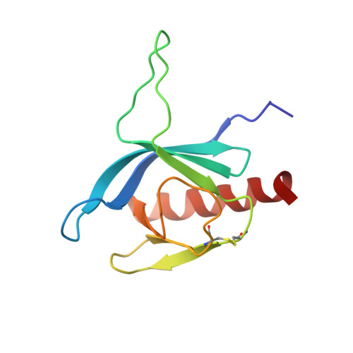
1P6S - PubMed Abstract:
The programmed cell death occurs as part of normal mammalian development. The induction of developmental cell death is a highly regulated process and can be suppressed by a variety of extracellular stimuli. Recently, the ability of trophic factors to promote survival have been attributed, at least in part, to the phosphatidylinositide 3'-OH kinase (PI3K)/Protein Kinase B (PKB, also named Akt) cascade. Several targets of the PI3K/PKB signaling pathway have been identified that may underlie the ability of this regulatory cascade to promote cell survival. PKB possesses a N-terminal Pleckstrin Homology (PH) domain that binds specifically and with high affinity to PtIns(3,4,5)P(3) and PtIns(3,4)P(2), the PI3K second messengers. PKB is then recruited to the plasma membrane by virtue of its interaction with 3'-OH phosphatidylinositides and activated. Recent evidence indicates that PKB is active in various types of human cancer; constitutive PKB signaling activation is believed to promote proliferation and increased cell survival, thereby contributing to cancer progression. Thus, it has been shown that induction of PKB activity is augmented by the TCL1/MTCP1 oncoproteins through a physical association requiring the PKB PH domain. Here we present the three-dimensional solution structure of the PH domain of the human protein PKB (isoform beta). PKBbeta-PH is an electrostatically polarized molecule that adopts the same fold and topology as other PH-domains, consisting of a beta-sandwich of seven strands capped on one top by an alpha-helix. The opposite face presents three variable loops that appear poorly defined in the NMR structure. Measurements of (15)N spin relaxation times and heteronuclear (15)N[(1)H]NOEs showed that this poor definition is due to intrinsic flexibility, involving complex motions on different time scales. Chemical shift mapping studies correctly defined the binding site of Ins(1,3,4,5)P(4) (the head group of PtIns(3,4,5)P(3)), as was previously proposed from a crystallographic study. More interestingly, these studies allowed us to define a putative alternative low-affinity binding site for Ins(1,4,5)P(3). The binding of this sugar to PKBbeta-PH might also involve non-specific association that could explain the stabilization of the protein in solution in the presence of Ins(1,4,5)P(3).
Organizational Affiliation:
Centre de Biochimie Structurale, UMR 5048 CNRS/UM1 - UMR 554 INSERM/UM1, Faculté de Pharmacie, BP14491, 15 Avenue Charles Flahault, 34093 Montpellier Cedex 5, France.