The 1.3-Angstrom-Resolution Crystal Structure of beta-Ketoacyl-Acyl Carrier Protein Synthase II from Streptococcus pneumoniae.
Price, A.C., Rock, C.O., White, S.W.(2003) J Bacteriol 185: 4136-4143
- PubMed: 12837788
- DOI: https://doi.org/10.1128/JB.185.14.4136-4143.2003
- Primary Citation of Related Structures:
1OX0, 1OXH - PubMed Abstract:
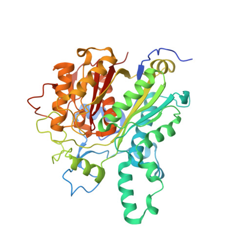
The beta-ketoacyl-acyl carrier protein synthases are members of the thiolase superfamily and are key regulators of bacterial fatty acid synthesis. As essential components of the bacterial lipid metabolic pathway, they are an attractive target for antibacterial drug discovery. We have determined the 1.3 A resolution crystal structure of the beta-ketoacyl-acyl carrier protein synthase II (FabF) from the pathogenic organism Streptococcus pneumoniae. The protein adopts a duplicated betaalphabetaalphabetaalphabetabeta fold, which is characteristic of the thiolase superfamily. The two-fold pseudosymmetry is broken by the presence of distinct insertions in the two halves of the protein. These insertions have evolved to bind the specific substrates of this particular member of the thiolase superfamily. Docking of the pantetheine moiety of the substrate identifies the loop regions involved in substrate binding and indicates roles for specific, conserved residues in the substrate binding tunnel. The active site triad of this superfamily is present in spFabF as His 303, His 337, and Cys 164. Near the active site is an ion pair, Glu 346 and Lys 332, that is conserved in the condensing enzymes but is unusual in our structure in being stabilized by an Mg(2+) ion which interacts with Glu 346. The active site histidines interact asymmetrically with Lys 332, whose positive charge is closer to His 303, and we propose a specific role for the lysine in polarizing the imidazole ring of this histidine. This asymmetry suggests that the two histidines have unequal roles in catalysis and provides new insights into the catalytic mechanisms of these enzymes.
Organizational Affiliation:
Department of Structural Biology, St. Jude Children's Research Hospital, Department of Molecular Sciences, University of Tennessee, Memphis, Tennessee 38105, USA.