1OW4

Crystal structure of a pheromone binding protein from the cockroach Leucophaea maderae in complex with the fluorescent reporter ANS (1-anilinonaphtalene-8-sulfonic acid),


Experimental Data Snapshot

  • Method: X-RAY DIFFRACTION
  • Resolution: 1.60 Å
  • R-Value Free: 0.208 
  • R-Value Work: 0.174 
  • R-Value Observed: 0.176 

wwPDB Validation   3D Report Full Report


Ligand Structure Quality Assessment 


This is version 1.3 of the entry. See complete history


Literature

THE CRYSTAL STRUCTURE OF A COCKROACH PHEROMONE-BINDING PROTEIN SUGGESTS A NEW LIGAND BINDING AND RELEASE MECHANISM

Lartigue, A.Gruez, A.Spinelli, S.Riviere, S.Brossut, R.Tegoni, M.Cambillau, C.

(2003) J Biol Chem 278: 30213-30218

  • DOI: https://doi.org/10.1074/jbc.M304688200
  • Primary Citation of Related Structures:  
    1ORG, 1OW4, 1P28

  • PubMed Abstract: 

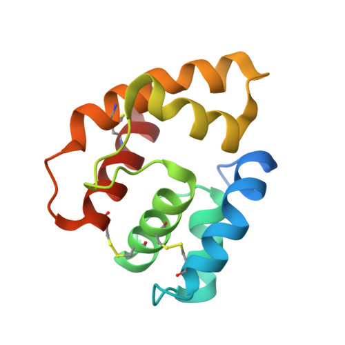
    Pheromone-binding proteins (PBPs) are small helical proteins found in sensorial organs, particularly in the antennae, of moth and other insect species. They were proposed to solubilize and carry the hydrophobic pheromonal compounds through the antennal lymph to receptors, participating thus in the peri-receptor events of signal transduction. The x-ray structure of Bombyx mori PBP (BmorPBP), from male antennae, revealed a six-helix fold forming a cavity that contains the pheromone bombykol. We have identified a PBP (LmaPBP) from the cockroach Leucophaea maderae in the antennae of the females, the gender attracted by pheromones in this species. Here we report the crystal structure of LmaPBP alone or in complex with a fluorescent reporter (amino-naphthalen sulfonate, ANS) or with a component of the pheromonal blend, 3-hydroxy-butan-2-one. Both compounds bind in the internal cavity of LmaPBP, which is more hydrophilic than BmorPBP cavity. LmaPBP structure ends just after the sixth helix (helix F). BmorPBP structure extends beyond the sixth helix with a stretch of residues elongated at neutral pH and folding as a seventh internalized helix at low pH. These differences between LmaPBP and BmorPBP structures suggest that different binding and release mechanism may be adapted to the hydrophilicity or hydrophobicity of the pheromonal ligand.


  • Organizational Affiliation

    Architecture et Fonction des Macromolécules Biologiques, UMR 6098 CNRS and Universités Aix-Marseille I & 2, 31 Ch. Joseph Aiguier, 13402 Marseille Cedex 20, France.


Macromolecules
Find similar proteins by:  (by identity cutoff)  |  3D Structure
Entity ID: 1
MoleculeChains Sequence LengthOrganismDetailsImage
pheromone binding protein
A, B
129Rhyparobia maderaeMutation(s): 0 
Gene Names: Pheromone Binding Protein (PBP)
Membrane Entity: Yes 
UniProt
Find proteins for Q8MTC1 (Rhyparobia maderae)
Explore Q8MTC1 
Go to UniProtKB:  Q8MTC1
Entity Groups  
Sequence Clusters30% Identity50% Identity70% Identity90% Identity95% Identity100% Identity
UniProt GroupQ8MTC1
Sequence Annotations
Expand
  • Reference Sequence
Small Molecules
Binding Affinity Annotations 
IDSourceBinding Affinity
2AN PDBBind:  1OW4 Kd: 2100 (nM) from 1 assay(s)
Binding MOAD:  1OW4 Kd: 2100 (nM) from 1 assay(s)
Experimental Data & Validation

Experimental Data

  • Method: X-RAY DIFFRACTION
  • Resolution: 1.60 Å
  • R-Value Free: 0.208 
  • R-Value Work: 0.174 
  • R-Value Observed: 0.176 
  • Space Group: P 1
Unit Cell:
Length ( Å )Angle ( ˚ )
a = 43.422α = 118.55
b = 45.07β = 92.63
c = 45.379γ = 107.17
Software Package:
Software NamePurpose
REFMACrefinement
DENZOdata reduction
CCP4data scaling
AMoREphasing

Structure Validation

View Full Validation Report


Ligand Structure Quality Assessment 


Entry History 

Deposition Data

Revision History  (Full details and data files)

  • Version 1.0: 2003-08-05
    Type: Initial release
  • Version 1.1: 2008-04-29
    Changes: Version format compliance
  • Version 1.2: 2011-07-13
    Changes: Advisory, Version format compliance
  • Version 1.3: 2024-04-03
    Changes: Data collection, Database references, Derived calculations, Refinement description