A Mechanism of Covalent Substrate Binding in the X-Ray Structure of Subunit K of the Escherichia Coli Dihydroxyacetone Kinase
Siebold, C., Garcia-Alles, L.-F., Erni, B., Baumann, U.(2003) Proc Natl Acad Sci U S A 100: 8188
- PubMed: 12813127
- DOI: https://doi.org/10.1073/pnas.0932787100
- Primary Citation of Related Structures:
1OI2, 1OI3 - PubMed Abstract:
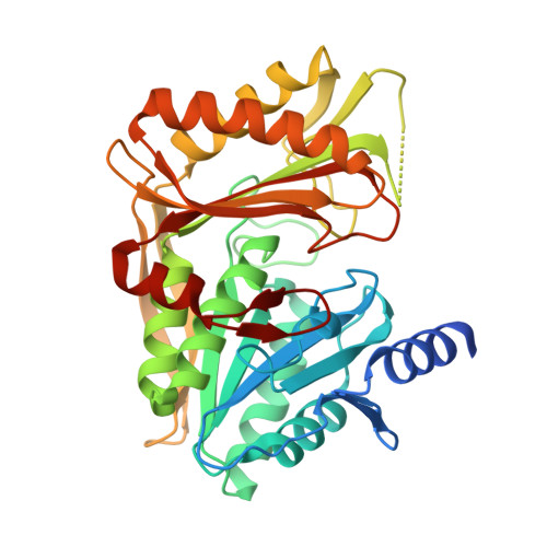
Dihydroxyacetone (Dha) kinases are homologous proteins that use different phosphoryl donors, a multiphosphoryl protein of the phosphoenolpyruvate-dependent carbohydrate:phosphotransferase system in bacteria, ATP in animals, plants, and some bacteria. The Dha kinase of Escherichia coli consists of three subunits, DhaK and DhaL, which are colinear to the ATP-dependent Dha kinases of eukaryotes, and the multiphosphoryl protein DhaM. Here we show the crystal structure of the DhaK subunit in complex with Dha at 1.75 A resolution. DhaK is a homodimer with a fold consisting of two six-stranded mixed beta-sheets surrounded by nine alpha-helices and a beta-ribbon covering the exposed edge strand of one sheet. The core of the N-terminal domain has an alpha/beta fold common to subunits of carbohydrate transporters and transcription regulators of the phosphoenolpyruvate-dependent carbohydrate:phosphotransferase system. The core of the C-terminal domain has a fold similar to the C-terminal domain of the cell-division protein FtsZ. A molecule of Dha is covalently bound in hemiaminal linkage to the N epsilon 2 of His-230. The hemiaminal does not participate in covalent catalysis but is the chemical basis for discrimination between short-chain carbonyl compounds and polyols. Paralogs of Dha kinases occur in association with transcription regulators of the TetR/QacR and the SorC families, pointing to their biological role as sensors in signaling.
Organizational Affiliation:
Departement für Chemie und Biochemie, Universität Bern, Freiestrasse 3, CH-3012 Bern, Switzerland.