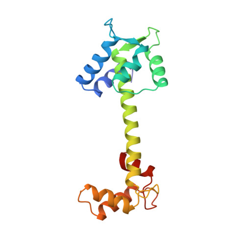
X-ray structures of Mn, Cd and Tb metal complexes of troponin C.
Rao, S.T., Satyshur, K.A., Greaser, M.L., Sundaralingam, M.(1996) Acta Crystallogr D Biol Crystallogr 52: 916-922
- PubMed: 15299599
- DOI: https://doi.org/10.1107/S0907444996006166
- Primary Citation of Related Structures:
1NCX, 1NCY, 1NCZ - PubMed Abstract:
The crystal structures of three metal complexes of troponin C (TnC) have been determined and refined where the two occupied structural Ca(2+) sites in the C domain have been substituted by Mn(2+), Cd(2+) and Tb(3+). The X-ray intensity data were collected to 2.1, 1.8 and 1.8 A resolution, respectively, on the three metal complexes, which are isomorphous with Ca-TnC. The three complexes have r.m.s. deviations of 0.27, 0.25 and 0.35 A, respectively, for all protein atoms, from Ca-TnC. Irrespective of the charge on the metal (+2 or +3), the occupied sites 3 and 4 exhibit a distorted pentagonal bipyramidal coordination, like Ca-TnC, with seven ligands, six from the 12-residue binding loop and the seventh from a water molecule. Mn(2+) at site 4 seems to display a longer distance to one of the carboxyl bidentate ligands representing an intermediate coordination simulating the six-coordinate Mg(2+). The carboxyl O atoms of the bidentate Glu12 are displaced on the side of the equatorial plane passing through the remaining three ligands with one O atom closer to the plane (Delta of 0.11 to 0.76 A) than the other (Delta of 0.93 to 1.38 A). The two axial ligands are an aspartic carboxyl O atom and a water molecule. The metal is displaced (0.18 to 0.56 A) towards the water facing the water channel.
Organizational Affiliation:
Department of Biochemistry, College of Agricultural & Life Sciences, University of Wisconsin-Madison, 53706, USA.