1N9K

Crystal structure of the bromide adduct of AphA class B acid phosphatase/phosphotransferase from E. coli at 2.2 A resolution


Experimental Data Snapshot

  • Method: X-RAY DIFFRACTION
  • Resolution: 2.20 Å
  • R-Value Free: 0.231 
  • R-Value Work: 0.189 
  • R-Value Observed: 0.191 

wwPDB Validation   3D Report Full Report


This is version 1.4 of the entry. See complete history


Literature

The first structure of a bacterial class B Acid phosphatase reveals further structural heterogeneity among phosphatases of the haloacid dehalogenase fold.

Calderone, V.Forleo, C.Benvenuti, M.Thaller, M.C.Rossolini, G.M.Mangani, S.

(2004) J Mol Biol 335: 761-773

  • DOI: https://doi.org/10.1016/j.jmb.2003.10.050
  • Primary Citation of Related Structures:  
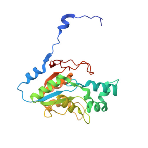
    1N8N, 1N9K

  • PubMed Abstract: 

    AphA is a periplasmic acid phosphatase of Escherichia coli belonging to class B bacterial phosphatases, which is part of the DDDD superfamily of phosphohydrolases. The crystal structure of AphA has been determined at 2.2A and its resolution extended to 1.7A on an AuCl(3) derivative. This represents the first crystal structure of a class B bacterial phosphatase. Despite the lack of sequence homology, the AphA structure reveals a haloacid dehalogenase-like fold. This finding suggests that this fold could be conserved among members of the DDDD superfamily of phosphohydrolases. The active enzyme is a homotetramer built by using an extended N-terminal arm intertwining the four monomers. The active site of the native enzyme, as prepared, hosts a magnesium ion, which can be replaced by other metal ions. The structure explains the non-specific behaviour of AphA towards substrates, while a structure-based alignment with other phosphatases provides clues about the catalytic mechanism.


  • Organizational Affiliation

    Dipartimento di Chimica, Università di Siena, Via Aldo Moro, I-53100 Siena, Italy.


Macromolecules
Find similar proteins by:  (by identity cutoff)  |  3D Structure
Entity ID: 1
MoleculeChains Sequence LengthOrganismDetailsImage
Class B acid phosphatase
A, B
212Escherichia coliMutation(s): 0 
Gene Names: APHA
EC: 3.1.3.2
UniProt
Find proteins for P0AE22 (Escherichia coli (strain K12))
Explore P0AE22 
Go to UniProtKB:  P0AE22
Entity Groups  
Sequence Clusters30% Identity50% Identity70% Identity90% Identity95% Identity100% Identity
UniProt GroupP0AE22
Sequence Annotations
Expand
  • Reference Sequence
Small Molecules
Ligands 2 Unique
IDChains Name / Formula / InChI Key2D Diagram3D Interactions
BR
Query on BR

Download Ideal Coordinates CCD File 
D [auth A]
E [auth A]
F [auth A]
G [auth A]
H [auth A]
D [auth A],
E [auth A],
F [auth A],
G [auth A],
H [auth A],
I [auth A],
J [auth A],
L [auth B],
M [auth B],
N [auth B],
O [auth B],
P [auth B],
Q [auth B],
R [auth B],
S [auth B]
BROMIDE ION
Br
CPELXLSAUQHCOX-UHFFFAOYSA-M
MG
Query on MG

Download Ideal Coordinates CCD File 
C [auth A],
K [auth B]
MAGNESIUM ION
Mg
JLVVSXFLKOJNIY-UHFFFAOYSA-N
Experimental Data & Validation

Experimental Data

  • Method: X-RAY DIFFRACTION
  • Resolution: 2.20 Å
  • R-Value Free: 0.231 
  • R-Value Work: 0.189 
  • R-Value Observed: 0.191 
  • Space Group: P 21 21 2
Unit Cell:
Length ( Å )Angle ( ˚ )
a = 49.499α = 90
b = 92.616β = 90
c = 138.247γ = 90
Software Package:
Software NamePurpose
DENZOdata reduction
SCALEPACKdata scaling
SOLVEphasing
RESOLVEmodel building
REFMACrefinement
RESOLVEphasing

Structure Validation

View Full Validation Report


Entry History 

Deposition Data

Revision History  (Full details and data files)

  • Version 1.0: 2004-02-03
    Type: Initial release
  • Version 1.1: 2008-04-28
    Changes: Version format compliance
  • Version 1.2: 2011-07-13
    Changes: Derived calculations, Version format compliance
  • Version 1.3: 2017-10-11
    Changes: Refinement description
  • Version 1.4: 2024-02-14
    Changes: Data collection, Database references, Derived calculations