Atomic structure of Mycobacterium tuberculosis CYP121 to 1.06 A reveals novel features of cytochrome P450.
Leys, D., Mowat, C.G., McLean, K.J., Richmond, A., Chapman, S.K., Walkinshaw, M.D., Munro, A.W.(2003) J Biol Chem 278: 5141-5147
- PubMed: 12435731
- DOI: https://doi.org/10.1074/jbc.M209928200
- Primary Citation of Related Structures:
1N40, 1N4G - PubMed Abstract:
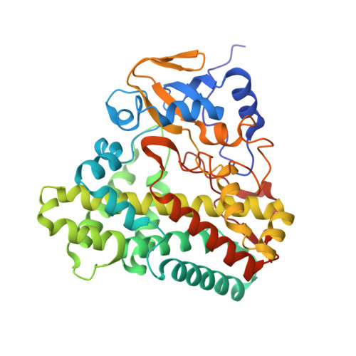
The first structure of a P450 to an atomic resolution of 1.06 A has been solved for CYP121 from Mycobacterium tuberculosis. A comparison with P450 EryF (CYP107A1) reveals a remarkable overall similarity in fold with major differences residing in active site structural elements. The high resolution obtained allows visualization of several unusual aspects. The heme cofactor is bound in two distinct conformations while being notably kinked in one pyrrole group due to close interaction with the proline residue (Pro(346)) immediately following the heme iron-ligating cysteine (Cys(345)). The active site is remarkably rigid in comparison with the remainder of the structure, notwithstanding the large cavity volume of 1350 A(3). The region immediately surrounding the distal water ligand is remarkable in several aspects. Unlike other bacterial P450s, the I helix shows no deformation, similar to mammalian CYP2C5. In addition, the positively charged Arg(386) is located immediately above the heme plane, dominating the local structure. Putative proton relay pathways from protein surface to heme (converging at Ser(279)) are identified. Most interestingly, the electron density indicates weak binding of a dioxygen molecule to the P450. This structure provides a basis for rational design of putative antimycobacterial agents.
Organizational Affiliation:
Department of Biochemistry, University of Leicester, The Adrian Building, University Road, Leicester LE1 7RH, United Kingdom. dl37@le.ac.uk