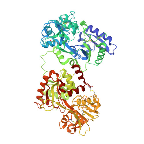
Structure of diferric hen serum transferrin at 2.8 A resolution.
Guha Thakurta, P., Choudhury, D., Dasgupta, R., Dattagupta, J.K.(2003) Acta Crystallogr D Biol Crystallogr 59: 1773-1781
- PubMed: 14501117
- DOI: https://doi.org/10.1107/s0907444903016652
- Primary Citation of Related Structures:
1N04 - PubMed Abstract:
Hen serum transferrin in its diferric form (hST) has been isolated, purified and the three-dimensional structure determined by X-ray crystallography at 2.8 A resolution. The final refined structure of hST, comprising 5232 protein atoms, two Fe(3+) cations, two CO(3)(2-) anions, 54 water molecules and one fucose moiety, has an R factor of 21.5% and an R(free) of 26.9% for all data. The structure has been compared with the three-dimensional structure of hen ovotransferrin (hOT) and also with structures of some other transferrins, viz. rabbit serum transferrin (rST) and human lactoferrin (hLF). The overall conformation of the hST molecule is essentially the same as that of other transferrins. However, the relative orientation of the two lobes, which is related to the species-specific receptor-recognition property of transferrins, has been found to be different in hST from that in hOT, rST and hLF. On the basis of superposition of the N lobes, rotations of 5.8, 16.9 and 11.3 degrees are required to bring the C lobes of hOT, rST and hLF, respectively, into coincidence with that of hST. A number of additional hydrogen bonds between the two domains in the N and C lobes have been identified in the structure of hST compared with that of hOT, which indicate a greater compactness of the lobes of hST than those of hOT. Being products of the same gene, hST and hOT have 100% sequence identity and differ only in the attached carbohydrate moiety. On the other hand, despite having similar functions, hST and rST have only 51% sequence similarity. However, the nature of the interdomain interactions of hST are closer to rST than to hOT. A putative carbohydrate-binding site has been identified in the N lobe of hST at Asn52 and a fucose molecule could be modelled at the site. The variations in interdomain and interlobe interactions in hST, together with altered lobe orientation with respect to hOT, rST and hLF, which are the representatives of the other subfamily of transferrins, are discussed.
Organizational Affiliation:
Crystallography and Molecular Biology Division, Saha Institute of Nuclear Physics, 1/AF Bidhannagar, Kolkata 700 064, India.