1MZ5

Trypanosoma rangeli sialidase


Experimental Data Snapshot

  • Method: X-RAY DIFFRACTION
  • Resolution: 2.20 Å
  • R-Value Free: 0.237 
  • R-Value Work: 0.180 
  • R-Value Observed: 0.183 

wwPDB Validation   3D Report Full Report


This is version 1.3 of the entry. See complete history


Literature

Structural basis of sialyltransferase activity in trypanosomal sialidases

Buschiazzo, A.Tavares, G.A.Campetella, O.Spinelli, S.Cremona, M.L.Paris, G.Amaya, M.F.Frasch, A.C.C.Alzari, P.M.

(2000) EMBO J 19: 16-24

  • DOI: https://doi.org/10.1093/emboj/19.1.16
  • Primary Citation of Related Structures:  
    1MZ5, 1MZ6

  • PubMed Abstract: 

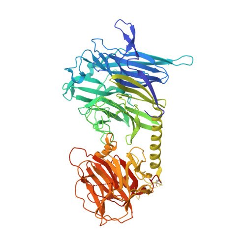
    The intracellular parasite Trypanosoma cruzi, the etiological agent of Chagas disease, sheds a developmentally regulated surface trans-sialidase, which is involved in key aspects of parasite-host cell interactions. Although it shares a common active site architecture with bacterial neuraminidases, the T.cruzi enzyme behaves as a highly efficient sialyltransferase. Here we report the crystal structure of the closely related Trypanosoma rangeli sialidase and its complex with inhibitor. The enzyme folds into two distinct domains: a catalytic beta-propeller fold tightly associated with a lectin-like domain. Comparison with the modeled structure of T.cruzi trans-sialidase and mutagenesis experiments allowed the identification of amino acid substitutions within the active site cleft that modulate sialyltransferase activity and suggest the presence of a distinct binding site for the acceptor carbohydrate. The structures of the Trypanosoma enzymes illustrate how a glycosidase scaffold can achieve efficient glycosyltransferase activity and provide a framework for structure-based drug design.


  • Organizational Affiliation

    Unité de Biochimie Structurale, Institut Pasteur, 25 rue du Dr Roux, 75724 Paris.


Macromolecules
Find similar proteins by:  (by identity cutoff)  |  3D Structure
Entity ID: 1
MoleculeChains Sequence LengthOrganismDetailsImage
sialidase638Trypanosoma rangeliMutation(s): 0 
EC: 3.2.1.18
UniProt
Find proteins for O44049 (Trypanosoma rangeli)
Explore O44049 
Go to UniProtKB:  O44049
Entity Groups  
Sequence Clusters30% Identity50% Identity70% Identity90% Identity95% Identity100% Identity
UniProt GroupO44049
Sequence Annotations
Expand
  • Reference Sequence
Experimental Data & Validation

Experimental Data

  • Method: X-RAY DIFFRACTION
  • Resolution: 2.20 Å
  • R-Value Free: 0.237 
  • R-Value Work: 0.180 
  • R-Value Observed: 0.183 
  • Space Group: P 21 21 21
Unit Cell:
Length ( Å )Angle ( ˚ )
a = 76.15α = 90
b = 93.41β = 90
c = 105.48γ = 90
Software Package:
Software NamePurpose
DENZOdata reduction
SCALEPACKdata scaling
AMoREphasing
REFMACrefinement
MLPHAREphasing

Structure Validation

View Full Validation Report


Entry History 

Deposition Data

Revision History  (Full details and data files)

  • Version 1.0: 2002-10-16
    Type: Initial release
  • Version 1.1: 2008-04-28
    Changes: Version format compliance
  • Version 1.2: 2011-07-13
    Changes: Non-polymer description, Version format compliance
  • Version 1.3: 2020-07-29
    Type: Remediation
    Reason: Carbohydrate remediation
    Changes: Data collection, Database references, Derived calculations, Structure summary