1MF0

Structure of the Recombinant Mouse-Muscle Adenylosuccinate Synthetase Complexed with AMP, GDP, HPO4(2-), and Mg(2+)


Experimental Data Snapshot

  • Method: X-RAY DIFFRACTION
  • Resolution: 2.50 Å
  • R-Value Free: 0.292 
  • R-Value Work: 0.219 

wwPDB Validation   3D Report Full Report


Ligand Structure Quality Assessment 


This is version 1.4 of the entry. See complete history


Literature

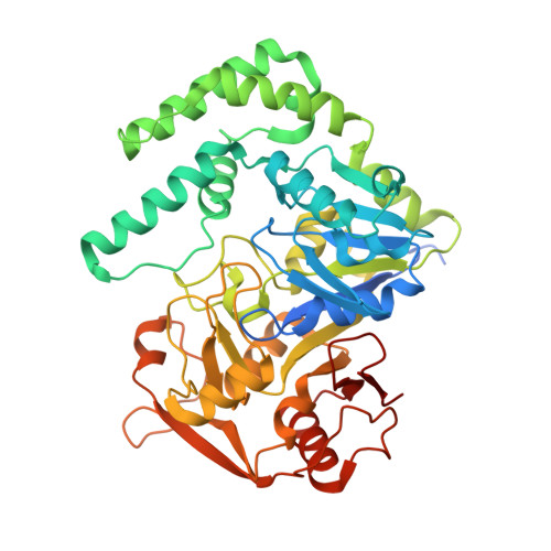
Feedback inhibition and product complexes of recombinant mouse muscle adenylosuccinate synthetase.

Iancu, C.V.Borza, T.Fromm, H.J.Honzatko, R.B.

(2002) J Biol Chem 277: 40536-40543

  • DOI: https://doi.org/10.1074/jbc.M204952200
  • Primary Citation of Related Structures:  
    1MEZ, 1MF0, 1MF1

  • PubMed Abstract: 

    Adenylosuccinate synthetase governs the committed step of AMP biosynthesis, the generation of 6-phosphoryl-IMP from GTP and IMP followed by the formation of adenylosuccinate from 6-phosphoryl-IMP and l-aspartate. The enzyme is subject to feedback inhibition by AMP and adenylosuccinate, but crystallographic complexes of the mouse muscle synthetase presented here infer mechanisms of inhibition that involve potentially synergistic ligand combinations. AMP alone adopts the productive binding mode of IMP and yet stabilizes the active site in a conformation that favors the binding of Mg(2+)-IMP to the GTP pocket. On the other hand, AMP, in the presence of GDP, orthophosphate, and Mg(2+), adopts the binding mode of adenylosuccinate. Depending on circumstances then, AMP behaves as an analogue of IMP or as an analogue of adenylosuccinate. The complex of adenylosuccinate.GDP.Mg(2+).sulfate, the first structure of an adenylosuccinate-bound synthetase, reveals significant geometric distortions and tight nonbonded contacts relevant to the proposed catalytic mechanism. Adenylosuccinate forms from 6-phosphoryl-IMP and l-aspartate by the movement of the purine ring into the alpha-amino group of l-aspartate.


  • Organizational Affiliation

    Department of Biochemistry, Biophysics, and Molecular Biology, Iowa State University, Ames, IA 50011, USA.


Macromolecules
Find similar proteins by:  (by identity cutoff)  |  3D Structure
Entity ID: 1
MoleculeChains Sequence LengthOrganismDetailsImage
Adenylosuccinate Synthetase457Mus musculusMutation(s): 0 
Gene Names: Adss1
EC: 6.3.4.4
UniProt
Find proteins for P28650 (Mus musculus)
Explore P28650 
Go to UniProtKB:  P28650
Entity Groups  
Sequence Clusters30% Identity50% Identity70% Identity90% Identity95% Identity100% Identity
UniProt GroupP28650
Sequence Annotations
Expand
  • Reference Sequence
Experimental Data & Validation

Experimental Data

  • Method: X-RAY DIFFRACTION
  • Resolution: 2.50 Å
  • R-Value Free: 0.292 
  • R-Value Work: 0.219 
  • Space Group: P 43 21 2
Unit Cell:
Length ( Å )Angle ( ˚ )
a = 70.6α = 90
b = 70.6β = 90
c = 198.39γ = 90
Software Package:
Software NamePurpose
AMoREphasing
CNSrefinement

Structure Validation

View Full Validation Report


Ligand Structure Quality Assessment 


Entry History 

Deposition Data

Revision History  (Full details and data files)

  • Version 1.0: 2002-10-30
    Type: Initial release
  • Version 1.1: 2008-04-28
    Changes: Version format compliance
  • Version 1.2: 2011-07-13
    Changes: Derived calculations, Version format compliance
  • Version 1.3: 2017-10-11
    Changes: Refinement description
  • Version 1.4: 2024-03-13
    Changes: Data collection, Database references, Derived calculations