1LQS

CRYSTAL STRUCTURE OF HUMAN CYTOMEGALOVIRUS IL-10 BOUND TO SOLUBLE HUMAN IL-10R1


Experimental Data Snapshot

  • Method: X-RAY DIFFRACTION
  • Resolution: 2.70 Å
  • R-Value Free: 0.294 
  • R-Value Work: 0.244 

wwPDB Validation   3D Report Full Report


Ligand Structure Quality Assessment 


This is version 1.5 of the entry. See complete history


Literature

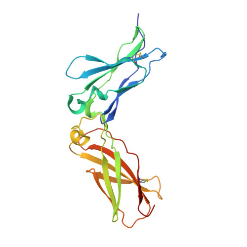
Crystal structure of human cytomegalovirus IL-10 bound to soluble human IL-10R1.

Jones, B.C.Logsdon, N.J.Josephson, K.Cook, J.Barry, P.A.Walter, M.R.

(2002) Proc Natl Acad Sci U S A 99: 9404-9409

  • DOI: https://doi.org/10.1073/pnas.152147499
  • Primary Citation of Related Structures:  
    1LQS

  • PubMed Abstract: 

    Human IL-10 (hIL-10) modulates critical immune and inflammatory responses by way of interactions with its high- (IL-10R1) and low-affinity (IL-10R2) cell surface receptors. Human cytomegalovirus exploits the IL-10 signaling pathway by expressing a functional viral IL-10 homolog (cmvIL-10), which shares only 27% sequence identity with hIL-10 yet signals through IL-10R1 and IL-10R2. To define the molecular basis of this virus-host interaction, we determined the 2.7-A crystal structure of cmvIL-10 bound to the extracellular fragment of IL-10R1 (sIL-10R1). The structure reveals cmvIL-10 forms a disulfide-linked homodimer that binds two sIL-10R1 molecules. Although cmvIL-10 and hIL-10 share similar intertwined topologies and sIL-10R1 binding sites, their respective interdomain angles differ by approximately 40 degrees. This difference results in a striking re-organization of the IL-10R1s in the putative cell surface complex. Solution binding studies show cmvIL-10 and hIL-10 share essentially identical affinities for sIL-10R1 whereas the Epstein-Barr virus IL-10 homolog (ebvIL-10), whose structure is highly similar to hIL-10, exhibits a approximately 20-fold reduction in sIL-10R1 affinity. Our results suggest cmvIL-10 and ebvIL-10 have evolved different molecular mechanisms to engage the IL-10 receptors that ultimately enhance the respective ability of their virus to escape immune detection.


  • Organizational Affiliation

    Center for Biophysical Sciences and Engineering, Department of Microbiology, University of Alabama, 1025 18th Street South, Birmingham, AL 35294, USA.


Macromolecules
Find similar proteins by:  (by identity cutoff)  |  3D Structure
Entity ID: 1
MoleculeChains Sequence LengthOrganismDetailsImage
INTERLEUKIN-10 RECEPTOR ALPHA CHAINA [auth R],
B [auth S]
214Homo sapiensMutation(s): 6 
UniProt & NIH Common Fund Data Resources
Find proteins for Q13651 (Homo sapiens)
Explore Q13651 
Go to UniProtKB:  Q13651
PHAROS:  Q13651
GTEx:  ENSG00000110324 
Entity Groups  
Sequence Clusters30% Identity50% Identity70% Identity90% Identity95% Identity100% Identity
UniProt GroupQ13651
Sequence Annotations
Expand
  • Reference Sequence
Find similar proteins by:  (by identity cutoff)  |  3D Structure
Entity ID: 2
MoleculeChains Sequence LengthOrganismDetailsImage
INTERLEUKIN-10-LIKE PROTEINC [auth L],
D [auth M]
157Human betaherpesvirus 5Mutation(s): 0 
UniProt
Find proteins for P17150 (Human cytomegalovirus (strain AD169))
Explore P17150 
Go to UniProtKB:  P17150
Entity Groups  
Sequence Clusters30% Identity50% Identity70% Identity90% Identity95% Identity100% Identity
UniProt GroupP17150
Sequence Annotations
Expand
  • Reference Sequence
Small Molecules
Ligands 1 Unique
IDChains Name / Formula / InChI Key2D Diagram3D Interactions
NAG
Query on NAG

Download Ideal Coordinates CCD File 
E [auth L],
F [auth M]
2-acetamido-2-deoxy-beta-D-glucopyranose
C8 H15 N O6
OVRNDRQMDRJTHS-FMDGEEDCSA-N
Experimental Data & Validation

Experimental Data

  • Method: X-RAY DIFFRACTION
  • Resolution: 2.70 Å
  • R-Value Free: 0.294 
  • R-Value Work: 0.244 
  • Space Group: C 1 2 1
Unit Cell:
Length ( Å )Angle ( ˚ )
a = 114.29α = 90
b = 104.68β = 106.49
c = 91.89γ = 90
Software Package:
Software NamePurpose
SCALEPACKdata scaling
CNSrefinement
CNSphasing

Structure Validation

View Full Validation Report


Ligand Structure Quality Assessment 


Entry History 

Deposition Data

Revision History  (Full details and data files)

  • Version 1.0: 2002-07-17
    Type: Initial release
  • Version 1.1: 2008-04-28
    Changes: Version format compliance
  • Version 1.2: 2011-07-13
    Changes: Non-polymer description, Version format compliance
  • Version 1.3: 2017-10-11
    Changes: Refinement description
  • Version 1.4: 2020-07-29
    Type: Remediation
    Reason: Carbohydrate remediation
    Changes: Data collection, Derived calculations, Structure summary
  • Version 1.5: 2021-10-27
    Changes: Database references, Structure summary