1LOA

THREE-DIMENSIONAL STRUCTURES OF COMPLEXES OF LATHYRUS OCHRUS ISOLECTIN I WITH GLUCOSE AND MANNOSE: FINE SPECIFICITY OF THE MONOSACCHARIDE-BINDING SITE


Experimental Data Snapshot

  • Method: X-RAY DIFFRACTION
  • Resolution: 2.20 Å
  • R-Value Work: 0.179 
  • R-Value Observed: 0.179 

wwPDB Validation   3D Report Full Report


Ligand Structure Quality Assessment 


This is version 1.4 of the entry. See complete history


Literature

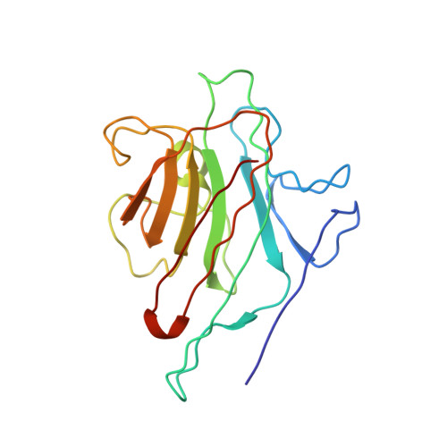
Three-dimensional structures of complexes of Lathyrus ochrus isolectin I with glucose and mannose: fine specificity of the monosaccharide-binding site.

Bourne, Y.Roussel, A.Frey, M.Rouge, P.Fontecilla-Camps, J.C.Cambillau, C.

(1990) Proteins 8: 365-376

  • DOI: https://doi.org/10.1002/prot.340080410
  • Primary Citation of Related Structures:  
    1LOA, 1LOB

  • PubMed Abstract: 

    The structure of the methyl-alpha-D-mannopyranoside-LOL I complex has been solved by the molecular replacement method using the refined saccharide-free LOL I coordinates as starting model. The methyl-alpha-D-mannopyranoside-LOL I complex was refined by simulated annealing using the program X-PLOR. The final R-factor value is 0.182 [Fo greater than 1 sigma(Fo)]. The isostructural methyl-alpha-D-glucopyranoside-LOL I complex was refined by X-Ray coupled energy minimization using the methyl-alpha-D-mannopyranoside-LOL I structure as a starting model to an R factor of 0.179 (all data). In both crystal forms, each dimer binds two molecules of sugar in pockets found near the calcium ions. The two saccharide moieties, which are in the C1 chair conformation, establish the same hydrogen bond pattern with the lectin. However, the van der Waals contacts are different between the O2, C2, C6, and O6 atoms of the two molecules and the backbone atoms of residues 208-211. Mannose, due to its axial C2 conformation, encloses the backbone atoms of the protein in a clamplike way. Van der Waals energy calculations suggest that this better complementarity of the mannoside molecule with the lectin could explain its higher affinity for isolectin I.


  • Organizational Affiliation

    Laboratoire de Cristallographie et de Cristallisation des Macromolécules Biologiques, Faculté de Médecine, Marseille, France.


Macromolecules
Find similar proteins by:  (by identity cutoff)  |  3D Structure
Entity ID: 1
MoleculeChains Sequence LengthOrganismDetailsImage
LEGUME ISOLECTIN I (ALPHA CHAIN)
A, C, E, G
181Lathyrus ochrusMutation(s): 0 
UniProt
Find proteins for P04122 (Lathyrus ochrus)
Explore P04122 
Go to UniProtKB:  P04122
Entity Groups  
Sequence Clusters30% Identity50% Identity70% Identity90% Identity95% Identity100% Identity
UniProt GroupP04122
Sequence Annotations
Expand
  • Reference Sequence
Find similar proteins by:  (by identity cutoff)  |  3D Structure
Entity ID: 2
MoleculeChains Sequence LengthOrganismDetailsImage
LEGUME ISOLECTIN I (BETA CHAIN)
B, D, F, H
52Lathyrus ochrusMutation(s): 0 
UniProt
Find proteins for P12306 (Lathyrus ochrus)
Explore P12306 
Go to UniProtKB:  P12306
Entity Groups  
Sequence Clusters30% Identity50% Identity70% Identity90% Identity95% Identity100% Identity
UniProt GroupP12306
Sequence Annotations
Expand
  • Reference Sequence
Small Molecules
Ligands 3 Unique
IDChains Name / Formula / InChI Key2D Diagram3D Interactions
GYP
Query on GYP

Download Ideal Coordinates CCD File 
I [auth A],
L [auth C],
O [auth E],
R [auth G]
methyl alpha-D-glucopyranoside
C7 H14 O6
HOVAGTYPODGVJG-ZFYZTMLRSA-N
MN
Query on MN

Download Ideal Coordinates CCD File 
K [auth A],
N [auth C],
Q [auth F],
T [auth G]
MANGANESE (II) ION
Mn
WAEMQWOKJMHJLA-UHFFFAOYSA-N
CA
Query on CA

Download Ideal Coordinates CCD File 
J [auth A],
M [auth C],
P [auth E],
S [auth G]
CALCIUM ION
Ca
BHPQYMZQTOCNFJ-UHFFFAOYSA-N
Experimental Data & Validation

Experimental Data

  • Method: X-RAY DIFFRACTION
  • Resolution: 2.20 Å
  • R-Value Work: 0.179 
  • R-Value Observed: 0.179 
  • Space Group: P 1 21 1
Unit Cell:
Length ( Å )Angle ( ˚ )
a = 56.3α = 90
b = 139.8β = 91
c = 62.7γ = 90
Software Package:
Software NamePurpose
X-PLORmodel building
X-PLORrefinement
X-PLORphasing

Structure Validation

View Full Validation Report


Ligand Structure Quality Assessment 


Entry History 

Deposition Data

Revision History  (Full details and data files)

  • Version 1.0: 1994-04-30
    Type: Initial release
  • Version 1.1: 2008-03-03
    Changes: Version format compliance
  • Version 1.2: 2011-07-13
    Changes: Version format compliance
  • Version 1.3: 2020-07-29
    Type: Remediation
    Reason: Carbohydrate remediation
    Changes: Data collection, Database references, Derived calculations, Other, Structure summary
  • Version 1.4: 2024-02-14
    Changes: Data collection, Database references, Structure summary