Three-dimensional structure of a cysteine-rich repeat from the low-density lipoprotein receptor.
Daly, N.L., Scanlon, M.J., Djordjevic, J.T., Kroon, P.A., Smith, R.(1995) Proc Natl Acad Sci U S A 92: 6334-6338
- PubMed: 7603991
- DOI: https://doi.org/10.1073/pnas.92.14.6334
- Primary Citation of Related Structures:
1LDL - PubMed Abstract:
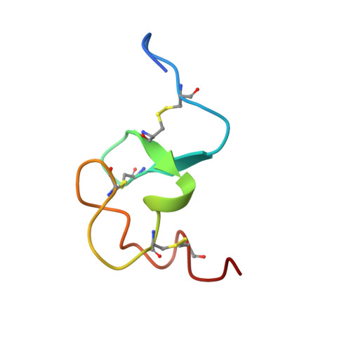
The low-density lipoprotein (LDL) receptor plays a central role in mammalian cholesterol metabolism, clearing lipoproteins which bear apolipoproteins E and B-100 from plasma. Mutations in this molecule are associated with familial hypercholesterolemia, a condition which leads to an elevated plasma cholesterol concentration and accelerated atherosclerosis. The N-terminal segment of the LDL receptor contains a heptad of cysteine-rich repeats that bind the lipoproteins. Similar repeats are present in related receptors, including the very low-density lipoprotein receptor and the LDL receptor-related protein/alpha 2-macroglobulin receptor, and in proteins which are functionally unrelated, such as the C9 component of complement. The first repeat of the human LDL receptor has been expressed in Escherichia coli as a glutathione S-transferase fusion protein, and the cleaved and purified receptor module has been shown to fold to a single, fully oxidized form that is recognized by the monoclonal antibody IgG-C7 in the presence of calcium ions. The three-dimensional structure of this module has been determined by two-dimensional NMR spectroscopy and shown to consist of a beta-hairpin structure, followed by a series of beta turns. Many of the side chains of the acidic residues, including the highly conserved Ser-Asp-Glu triad, are clustered on one face of the module. To our knowledge, this structure has not previously been described in any other protein and may represent a structural paradigm both for the other modules in the LDL receptor and for the homologous domains of several other proteins. Calcium ions had only minor effects on the CD spectrum and no effect on the 1H NMR spectrum of the repeat, suggesting that they induce no significant conformational change.
Organizational Affiliation:
Biochemistry Department, University of Queensland, Australia.