1KQ2

Crystal Structure of an Hfq-RNA Complex


Experimental Data Snapshot

  • Method: X-RAY DIFFRACTION
  • Resolution: 2.71 Å
  • R-Value Free: 0.266 
  • R-Value Work: 0.204 
  • R-Value Observed: 0.204 

wwPDB Validation   3D Report Full Report


This is version 1.3 of the entry. See complete history


Literature

Structures of the pleiotropic translational regulator Hfq and an Hfq-RNA complex: a bacterial Sm-like protein.

Schumacher, M.A.Pearson, R.F.Moller, T.Valentin-Hansen, P.Brennan, R.G.

(2002) EMBO J 21: 3546-3556

  • DOI: https://doi.org/10.1093/emboj/cdf322
  • Primary Citation of Related Structures:  
    1KQ1, 1KQ2

  • PubMed Abstract: 

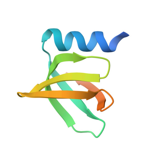
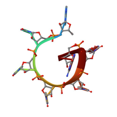
    In prokaryotes, Hfq regulates translation by modulating the structure of numerous RNA molecules by binding preferentially to A/U-rich sequences. To elucidate the mechanisms of target recognition and translation regulation by Hfq, we determined the crystal structures of the Staphylococcus aureus Hfq and an Hfq-RNA complex to 1.55 and 2.71 A resolution, respectively. The structures reveal that Hfq possesses the Sm-fold previously observed only in eukaryotes and archaea. However, unlike these heptameric Sm proteins, Hfq forms a homo-hexameric ring. The Hfq-RNA structure reveals that the single-stranded hepta-oligoribonucleotide binds in a circular conformation around a central basic cleft, whereby Tyr42 residues from adjacent subunits stack with six of the bases, and Gln8, outside the Sm motif, provides key protein-base contacts. Such binding suggests a mechanism for Hfq function.


  • Organizational Affiliation

    Department of Biochemistry and Molecular Biology, Oregon Health and Science University, Portland, OR 97201-3098, USA.


Macromolecules

Find similar proteins by:  (by identity cutoff)  |  3D Structure
Entity ID: 2
MoleculeChains Sequence LengthOrganismDetailsImage
Host factor for Q beta77Staphylococcus aureusMutation(s): 0 
UniProt
Find proteins for A0A0H3JV59 (Staphylococcus aureus (strain Mu50 / ATCC 700699))
Explore A0A0H3JV59 
Go to UniProtKB:  A0A0H3JV59
Entity Groups  
Sequence Clusters30% Identity50% Identity70% Identity90% Identity95% Identity100% Identity
UniProt GroupA0A0H3JV59
Sequence Annotations
Expand
  • Reference Sequence

Find similar nucleic acids by:  Sequence   |   3D Structure  

Entity ID: 1
MoleculeChains LengthOrganismImage
5'-R(*AP*UP*UP*UP*UP*UP*G)-3'A [auth R]7N/A
Sequence Annotations
Expand
  • Reference Sequence
Small Molecules
Binding Affinity Annotations 
IDSourceBinding Affinity
RNA PDBBind:  1KQ2 Kd: 30.5 (nM) from 1 assay(s)
Experimental Data & Validation

Experimental Data

  • Method: X-RAY DIFFRACTION
  • Resolution: 2.71 Å
  • R-Value Free: 0.266 
  • R-Value Work: 0.204 
  • R-Value Observed: 0.204 
  • Space Group: C 2 2 21
Unit Cell:
Length ( Å )Angle ( ˚ )
a = 80.87α = 90
b = 115.6β = 90
c = 101.84γ = 90
Software Package:
Software NamePurpose
MOLREPphasing
CNSrefinement
MOSFLMdata reduction
CCP4data scaling

Structure Validation

View Full Validation Report


Entry History 

Deposition Data

Revision History  (Full details and data files)

  • Version 1.0: 2002-07-05
    Type: Initial release
  • Version 1.1: 2008-04-27
    Changes: Version format compliance
  • Version 1.2: 2011-07-13
    Changes: Version format compliance
  • Version 1.3: 2023-08-16
    Changes: Data collection, Database references, Refinement description