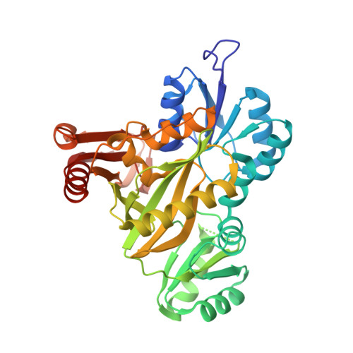
1KJ8

Crystal Structure of PurT-Encoded Glycinamide Ribonucleotide Transformylase in Complex with Mg-ATP and GAR


Experimental Data Snapshot

  • Method: X-RAY DIFFRACTION
  • Resolution: 1.60 Å
  • R-Value Free: 0.228 
  • R-Value Work: 0.181 
  • R-Value Observed: 0.182 

wwPDB Validation   3D Report Full Report


Ligand Structure Quality Assessment 


This is version 1.4 of the entry. See complete history


Literature

PurT-encoded glycinamide ribonucleotide transformylase. Accommodation of adenosine nucleotide analogs within the active site.

Thoden, J.B.Firestine, S.M.Benkovic, S.J.Holden, H.M.

(2002) J Biol Chem 277: 23898-23908

  • DOI: https://doi.org/10.1074/jbc.M202251200
  • Primary Citation of Related Structures:  
    1KJ8, 1KJ9, 1KJI, 1KJJ, 1KJQ

  • PubMed Abstract: 

    PurT-encoded glycinamide ribonucleotide transformylase, or PurT transformylase, functions in purine biosynthesis by catalyzing the formylation of glycinamide ribonucleotide through a catalytic mechanism requiring Mg(2+)ATP and formate. From previous x-ray diffraction analyses, it has been demonstrated that PurT transformylase from Escherichia coli belongs to the ATP-grasp superfamily of enzymes, which are characterized by three structural motifs referred to as the A-, B-, and C-domains. In all of the ATP-grasp enzymes studied to date, the adenosine nucleotide ligands are invariably wedged between the B- and C-domains, and in some cases, such as biotin carboxylase and carbamoyl phosphate synthetase, the B-domains move significantly upon nucleotide binding. Here we present a systematic and high-resolution structural investigation of PurT transformylase complexed with various adenosine nucleotides or nucleotide analogs including Mg(2+)ATP, Mg(2+)-5'-adenylylimidodiphosphate, Mg(2+)-beta,gamma-methyleneadenosine 5'-triphosphate, Mg(2+)ATPgammaS, or Mg(2+)ADP. Taken together, these studies indicate that the conformation of the so-called "T-loop," delineated by Lys-155 to Gln-165, is highly sensitive to the chemical identity of the nucleotide situated in the binding pocket. This sensitivity to nucleotide identity is in sharp contrast to that observed for the "P-loop"-containing enzymes, in which the conformation of the binding motif is virtually unchanged in the presence or absence of nucleotides.


  • Organizational Affiliation

    Department of Biochemistry, University of Wisconsin, Madison, Wisconsin 53706, USA.


Macromolecules
Find similar proteins by:  (by identity cutoff)  |  3D Structure
Entity ID: 1
MoleculeChains Sequence LengthOrganismDetailsImage
phosphoribosylglycinamide formyltransferase 2
A, B
391Escherichia coliMutation(s): 0 
Gene Names: PURT
EC: 2.1.2
UniProt
Find proteins for P33221 (Escherichia coli (strain K12))
Explore P33221 
Go to UniProtKB:  P33221
Entity Groups  
Sequence Clusters30% Identity50% Identity70% Identity90% Identity95% Identity100% Identity
UniProt GroupP33221
Sequence Annotations
Expand
  • Reference Sequence
Small Molecules
Ligands 7 Unique
IDChains Name / Formula / InChI Key2D Diagram3D Interactions
ATP
Query on ATP

Download Ideal Coordinates CCD File 
I [auth A],
Q [auth B]
ADENOSINE-5'-TRIPHOSPHATE
C10 H16 N5 O13 P3
ZKHQWZAMYRWXGA-KQYNXXCUSA-N
GAR
Query on GAR

Download Ideal Coordinates CCD File 
J [auth A],
R [auth B]
GLYCINAMIDE RIBONUCLEOTIDE
C7 H13 N2 O8 P
OBQMLSFOUZUIOB-SHUUEZRQSA-L
MPO
Query on MPO

Download Ideal Coordinates CCD File 
K [auth A]3[N-MORPHOLINO]PROPANE SULFONIC ACID
C7 H15 N O4 S
DVLFYONBTKHTER-UHFFFAOYSA-N
EDO
Query on EDO

Download Ideal Coordinates CCD File 
L [auth A],
S [auth B],
T [auth B]
1,2-ETHANEDIOL
C2 H6 O2
LYCAIKOWRPUZTN-UHFFFAOYSA-N
CL
Query on CL

Download Ideal Coordinates CCD File 
F [auth A],
G [auth A],
H [auth A],
P [auth B]
CHLORIDE ION
Cl
VEXZGXHMUGYJMC-UHFFFAOYSA-M
MG
Query on MG

Download Ideal Coordinates CCD File 
C [auth A],
D [auth A],
M [auth B],
N [auth B]
MAGNESIUM ION
Mg
JLVVSXFLKOJNIY-UHFFFAOYSA-N
NA
Query on NA

Download Ideal Coordinates CCD File 
E [auth A],
O [auth B]
SODIUM ION
Na
FKNQFGJONOIPTF-UHFFFAOYSA-N
Experimental Data & Validation

Experimental Data

  • Method: X-RAY DIFFRACTION
  • Resolution: 1.60 Å
  • R-Value Free: 0.228 
  • R-Value Work: 0.181 
  • R-Value Observed: 0.182 
  • Space Group: P 21 21 2
Unit Cell:
Length ( Å )Angle ( ˚ )
a = 62.1α = 90
b = 179.3β = 90
c = 75.8γ = 90
Software Package:
Software NamePurpose
TNTrefinement
FRAMBOdata collection
SAINTdata scaling
TNTphasing

Structure Validation

View Full Validation Report


Ligand Structure Quality Assessment 


Entry History 

Deposition Data

Revision History  (Full details and data files)

  • Version 1.0: 2002-06-28
    Type: Initial release
  • Version 1.1: 2008-04-27
    Changes: Version format compliance
  • Version 1.2: 2011-07-13
    Changes: Version format compliance
  • Version 1.3: 2017-10-11
    Changes: Refinement description
  • Version 1.4: 2023-08-16
    Changes: Data collection, Database references, Derived calculations, Refinement description