1K61

MATALPHA2 HOMEODOMAIN BOUND TO DNA


Experimental Data Snapshot

  • Method: X-RAY DIFFRACTION
  • Resolution: 2.10 Å
  • R-Value Free: 0.277 
  • R-Value Work: 0.221 
  • R-Value Observed: 0.221 

wwPDB Validation   3D Report Full Report


This is version 1.3 of the entry. See complete history


Literature

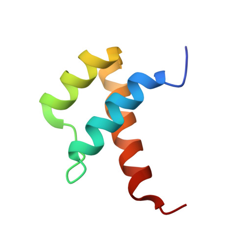
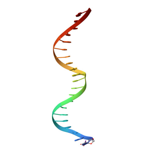
A Hoogsteen base pair embedded in undistorted B-DNA

Aishima, J.Gitti, R.K.Noah, J.E.Gan, H.H.Schlick, T.Wolberger, C.

(2002) Nucleic Acids Res 30: 5244-5252

  • DOI: https://doi.org/10.1093/nar/gkf661
  • Primary Citation of Related Structures:  
    1K61

  • PubMed Abstract: 

    Hoogsteen base pairs within duplex DNA typically are only observed in regions containing significant distortion or near sites of drug intercalation. We report here the observation of a Hoogsteen base pair embedded within undistorted, unmodified B-DNA. The Hoogsteen base pair, consisting of a syn adenine base paired with an anti thymine base, is found in the 2.1 A resolution structure of the MATalpha2 homeodomain bound to DNA in a region where a specifically and a non-specifically bound homeodomain contact overlapping sites. NMR studies of the free DNA show no evidence of Hoogsteen base pair formation, suggesting that protein binding favors the transition from a Watson-Crick to a Hoogsteen base pair. Molecular dynamics simulations of the homeodomain-DNA complex support a role for the non-specifically bound protein in favoring Hoogsteen base pair formation. The presence of a Hoogsteen base pair in the crystal structure of a protein-DNA complex raises the possibility that Hoogsteen base pairs could occur within duplex DNA and play a hitherto unrecognized role in transcription, replication and other cellular processes.


  • Organizational Affiliation

    Department of Biophysics and Biophysical Chemistry, Johns Hopkins University School of Medicine, Baltimore, MD 21205-2185, USA.


Macromolecules

Find similar proteins by:  (by identity cutoff)  |  3D Structure
Entity ID: 3
MoleculeChains Sequence LengthOrganismDetailsImage
Mating-type protein alpha-2C [auth A],
D [auth B],
E [auth C],
F [auth D]
60N/AMutation(s): 0 
UniProt
Find proteins for P0CY08 (Saccharomyces cerevisiae (strain ATCC 204508 / S288c))
Explore P0CY08 
Go to UniProtKB:  P0CY08
Entity Groups  
Sequence Clusters30% Identity50% Identity70% Identity90% Identity95% Identity100% Identity
UniProt GroupP0CY08
Sequence Annotations
Expand
  • Reference Sequence

Find similar nucleic acids by:  Sequence   |   3D Structure  

Entity ID: 1
MoleculeChains LengthOrganismImage
5'-D(*AP*CP*AP*TP*GP*TP*AP*AP*TP*TP*CP*AP*TP*TP*TP*AP*CP*AP*CP*GP*C)-3'A [auth E]21N/A
Sequence Annotations
Expand
  • Reference Sequence

Find similar nucleic acids by:  Sequence   |   3D Structure  

Entity ID: 2
MoleculeChains LengthOrganismImage
5'-D(*(5IU)P*GP*CP*GP*TP*GP*TP*AP*AP*AP*TP*GP*AP*AP*TP*TP*AP*CP*AP*TP*G)-3'B [auth F]21N/A
Sequence Annotations
Expand
  • Reference Sequence
Experimental Data & Validation

Experimental Data

  • Method: X-RAY DIFFRACTION
  • Resolution: 2.10 Å
  • R-Value Free: 0.277 
  • R-Value Work: 0.221 
  • R-Value Observed: 0.221 
  • Space Group: P 1 21 1
Unit Cell:
Length ( Å )Angle ( ˚ )
a = 38.94α = 90
b = 70.24β = 105.42
c = 68.29γ = 90
Software Package:
Software NamePurpose
SCALEPACKdata scaling
AMoREphasing
CNSrefinement

Structure Validation

View Full Validation Report


Entry History 

Deposition Data

Revision History  (Full details and data files)

  • Version 1.0: 2002-12-11
    Type: Initial release
  • Version 1.1: 2008-04-27
    Changes: Version format compliance
  • Version 1.2: 2011-07-13
    Changes: Version format compliance
  • Version 1.3: 2023-08-16
    Changes: Data collection, Database references, Derived calculations, Refinement description